
SLIDE 1
5/1/2009 1
Do we need biopsy to treat early inoperable lung cancer?
Frank J. Lagerwaard MD PhD VU University medical center Amsterdam, The Netherlands
VUmc Amsterdam Perspectives in Lung Cancer, Bruxelles, 2009
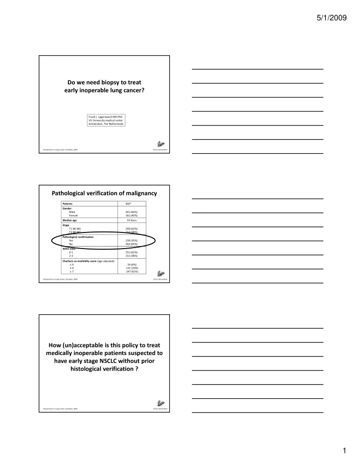
Pathological verification of malignancy
Patients 402* Gender Male Female 241 (60%) 161 (40%) Median age 74 Years Stage T1 N0 M0 T2 N0 M0 249 (62%) 153 (38%)
VUmc Amsterdam Perspectives in Lung Cancer, Bruxelles, 2009
Pathological confirmation Yes No 139 (35%) 263 (65%) WHO class 0‐1 2‐3 251 (62%) 151 (38%) Charlson co‐morbidity score (age‐adjusted) ≤ 4 5‐6 ≥ 7 24 (6%) 131 (33%) 247 (61%)
How (un)acceptable is this policy to treat medically inoperable patients suspected to have early stage NSCLC without prior
VUmc Amsterdam Perspectives in Lung Cancer, Bruxelles, 2009