3/13/2018 18 th Multidisciplinary Management of Cancers: A Casebased - - PDF document

3 13 2018
SMART_READER_LITE
LIVE PREVIEW

3/13/2018 18 th Multidisciplinary Management of Cancers: A Casebased - - PDF document

3/13/2018 18 th Multidisciplinary Management of Cancers: A Casebased Approach 18 th Multidisciplinary Management of Cancers: A Casebased Approach Matthew Gubens, MD MS, UC San Francisco, Chair Julia Rotow, MD, UC San Francisco, Fellow


slide-1
SLIDE 1

3/13/2018 1

18th Multidisciplinary Management of Cancers: A Case‐based Approach Multidisciplinary Management of Cancers Thoracic Oncology Tumor Board 18th Multidisciplinary Management of Cancers: A Case‐based Approach

Matthew Gubens, MD MS, UC San Francisco, Chair Julia Rotow, MD, UC San Francisco, Fellow Colin Blakely, MD PhD, UC San Francisco GigI Chen, MD, Diablo Valley Oncology & Hematology Medical Group David Cooke, MD, UC Davis Megan Daly, MD, UC Davis Maximilian Diehn, MD, Stanford David Gandara, MD, UC Davis Natalie Lui, MD, Stanford Johannes Kratz, MD, UC San Francisco Caroline McCoach, MD, UC San Francisco Jennifer Marie Suga, MD, The Permanente Medical Group Heather Wakelee, MD, Stanford

18th Multidisciplinary Management of Cancers: A Case‐based Approach

Case 1

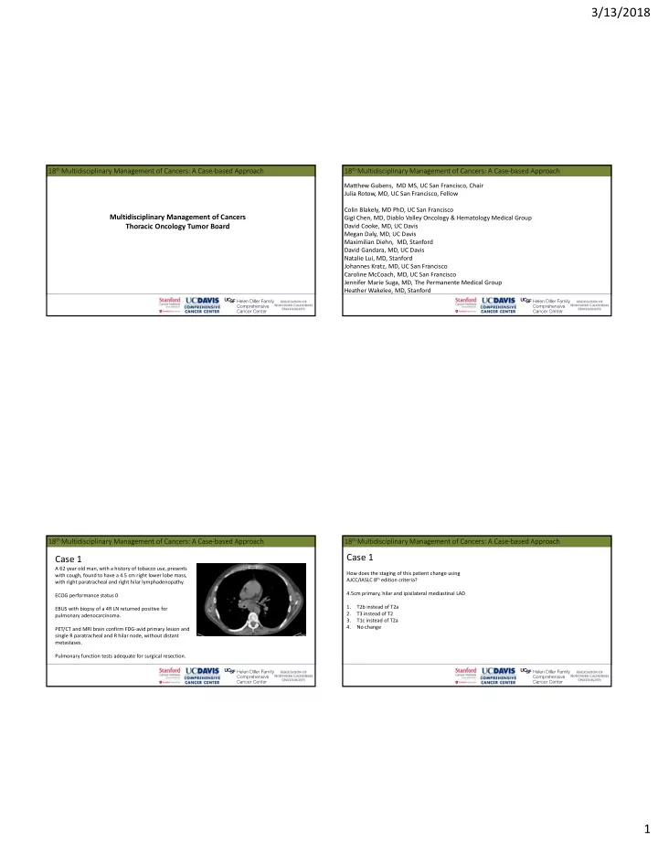
A 62 year old man, with a history of tobacco use, presents with cough, found to have a 4.5 cm right lower lobe mass, with right paratracheal and right hilar lymphadenopathy. ECOG performance status 0 EBUS with biopsy of a 4R LN returned positive for pulmonary adenocarcinoma. PET/CT and MRI brain confirm FDG‐avid primary lesion and single R paratracheal and R hilar node, without distant metastases. Pulmonary function tests adequate for surgical resection.

18th Multidisciplinary Management of Cancers: A Case‐based Approach

Case 1

How does the staging of this patient change using AJCC/IASLC 8th edition criteria? 4.5cm primary, hilar and ipisilateral mediastinal LAD 1. T2b instead of T2a 2. T3 instead of T2 3. T1c instead of T2a 4. No change

slide-2
SLIDE 2

3/13/2018 2

18th Multidisciplinary Management of Cancers: A Case‐based Approach 18th Multidisciplinary Management of Cancers: A Case‐based Approach 18th Multidisciplinary Management of Cancers: A Case‐based Approach

Case 1

What initial treatment strategy would you offer this patient with Stage IIIA (T2bN2) pulmonary adenocarcinoma? 1. Induction chemotherapy, followed by re‐evaluation for surgical resection 2. Induction chemoradiation, followed by re‐evaluation for surgical resection 3. Definitive chemoradiation with weekly carboplatin/paclitaxel 4. Definitive chemoradiation with cisplatin/etoposide 5. Definitive chemoradiation with cisplatin/pemetrexed

18th Multidisciplinary Management of Cancers: A Case‐based Approach

Case 1

The patient completes chemoradiation with good tolerance to therapy, PS1 at completion, with expected treatment effect without evidence of new or progressive disease at initial imaging. Now what do you recommend? 1. Begin surveillance with CT chest every 3‐6 months for the first 3 years, followed by less frequent surveillance intervals 2. Begin surveillance with CT chest/abdomen/pelvis every 3‐6 months for the first 3 years, followed by less frequent surveillance 3. Offer consolidation therapy with durvalumab (every two weeks) for up to one year 4. Offer molecular and PDL1 testing on diagnostic biopsy specimen to guide selection of consolidation therapy

slide-3
SLIDE 3

3/13/2018 3

PACIFIC Trial

Paz‐Ares, ESMO 2017

PACIFIC Trial

Paz‐Ares, ESMO 2017

PACIFIC Trial

Paz‐Ares, ESMO 2017

18th Multidisciplinary Management of Cancers: A Case‐based Approach

Case 1

What if the 4R LN from EBUS initially returned negative on

  • pathology. Right hilar LN sampling positive. PFTs

demonstrate a mild obstructive defect. How would you initially manage this T2bN1M0 pulmonary adenocarcinoma? 1. Additional mediastinoscopy for mediastinal LN staging 2. VATs resection of RUL with mediastinal LN dissection 3. Induction chemotherapy prior to consideration for resection

slide-4
SLIDE 4

3/13/2018 4

18th Multidisciplinary Management of Cancers: A Case‐based Approach

Case 1

The patient with clinical stage IIB (T2bN1M0) NSCLC undergoes a VATS right upper lobe lobectomy and mediastinal LN dissection. Pathology reveals a 3.9 cm pulmonary adenocarcinoma, R hilar node (1/10 nodes overall), margins positive (microscopic). What subsequent management would you offer to this patient with pT2aN1 adenocarcinoma. A) Surveillance every 3‐6 months with CT chest for the first year, then with decreasing frequency B) Concurrent chemoradiation C) Platinum doublet followed by radiation D) Offer re‐resection, followed by platinum doublet E) Chemoradiation, followed by consolidation with durvalumab

18th Multidisciplinary Management of Cancers: A Case‐based Approach

Case 1 Take Away Points

Consider consolidation durvalumab for patients with unresectable stage III NSCLC, following response to definitive chemoradiation. (NEJM 2017) The 8th edition of the AJCC guideline updates criteria for T and M staging.

18th Multidisciplinary Management of Cancers: A Case‐based Approach

Case 2

A 52 year old woman without co‐morbidities presents with a 7 cm left upper lobe mass, with mediastinal lymphadenopathy and bilateral pulmonary lesions up to 3 cm in size. Staging PET/CT and MRI brain also demonstrate 3 frontal lobe metastases up to 5 mm in size. The patient is asymptomatic from the brain mets, ECOG PS 0. Biopsy of the left upper lobe lesion shows pulmonary adenocarcinoma. Molecular testing is positive for EGFR L858R. PD‐L1 IHC (22C3): 60% positive tumor cells

18th Multidisciplinary Management of Cancers: A Case‐based Approach

Case 2

How would you treat this 52F with new diagnosis of stage IV, EGFR L858R+ adenocarcinoma with CNS involvement? 1. Refer for consideration for SBRT to CNS lesions, then start an oral EGFR TKI 2. Start with afatinib 3. Start with erlotinib 4. Start with gefitinib 5. Start with osimertinb

slide-5
SLIDE 5

3/13/2018 5

18th Multidisciplinary Management of Cancers: A Case‐based Approach

Case 2

The patient begins first‐line treatment with osimertinib at the usual 80 mg po daily dosing. She tolerates therapy well with minimal toxicity. CT chest/abdomen/pelvis obtained after the initial 2 months of treatment show a partial response, with approximately 50% decrease in pulmonary lesions and mediastinal adenopathy. MRI brain shows decrease in previously seen 3 frontal lobe lesions, largest 2 mm in size.

18th Multidisciplinary Management of Cancers: A Case‐based Approach

Case 2

After 12 months treatment with osimertinib, scans show the following compared to imaging 3 months prior: ‐‐1‐2 mm increase in a subcarinal LN and a 1‐2 mm increase in left upper lobe mass, currently 2 cm,

  • ther pulmonary nodule and lymph nodes remain stable.

‐‐In the CNS, one frontal lesion has increased in size from 2 mm to 1 cm with mild surrounding edema. The patient continues to feel well, without new pulmonary or neurologic symptoms.

18th Multidisciplinary Management of Cancers: A Case‐based Approach

Case 2

What is your treatment recommendation for this patient with EGFR‐mutant NSCLC with mild lung progression and new CNS disease during treatment with osimertinib? 1. Continue osimertinib, refer for consideration for SBRT, with plan for reimaging of systemic disease in 2‐3 months 2. Refer for consideration for SBRT and refer for biopsy of a progressing chest lesion for molecular testing. 3. Refer for consideration for SBRT and plan to change systemic therapy to platinum‐based chemotherapy 4. Change systemic therapy to platinum‐based chemotherapy

18th Multidisciplinary Management of Cancers: A Case‐based Approach

Case 2

After completing SBRT to three CNS lesions, the patient continues on osimertinib. Subsequent scans over the next six months show stability versus slow increase in her LUL mass and mediastinal adenopathy. Her most recent scans show a 1.5 cm increase in her LUL mass (now 3.5 cm in size) with newly enlarged left‐side retroperitoneal and left common iliac LN, up to approximately 3 cm in size. MRI brain without new or progressive disease. She is more fatigued and has begun to lose weight, ECOG PS 1.

slide-6
SLIDE 6

3/13/2018 6

18th Multidisciplinary Management of Cancers: A Case‐based Approach

Case 2

What is your treatment recommendation for this patient with EGFR‐mutant NSCLC with symptomatic systemic progression during treatment with osimertinib, with PD‐L1 60%? 1. Continue osimertinib but decrease imaging interval to 6 weeks 2. Obtain biopsy of an enlarging abdominal node with molecular and PD‐L1 testing 3. Discuss beginning treatment with pembrolizumab as next line of therapy 4. Discuss beginning treatment with carboplatin/pemetrexed/bevacizumab as next line of therapy 5. Discuss beginning treatment with carboplatin/pemetrexed/pembrolizumab as next line of therapy

Ramalingam S, et al. ESMO 2017. Abstr LBA2_PR.

FLAURA: Osimertinib vs. Gefitinib/Erlotinib in EGFR Mutated NSCLC

18th Multidisciplinary Management of Cancers: A Case‐based Approach

Case 2

What if this 52F with new diagnosis of stage IV EGFR L858R adenocarcinoma, but without CNS involvement, had instead begun first‐line treatment with erlotinib. She is tolerating treatment well, with response as of 6 months treatment. She asks if she should be taking osimertinib instead of erlotinib. You recommend: 1. Continue treatment with erlotinib. At progression, transition to osimertinib if EGFR T790M present. 2. Continue treatment with erlotinib. At progression transition to osimertinib without evaluation for T790M. 3. Switch to treatment with osimertinib now 4. Switch to osimertinib prior to progression only if intolerant to erlotinib.

18th Multidisciplinary Management of Cancers: A Case‐based Approach

Case 2

You continue erlotinib in if this 52F with stage IV EGFR L858R adenocarcinoma. After 4 more months, there is evidence of progressive disease in multiple lung nodules. You decide to test for the presence of the T790M resistance mutation. How would you test for T790M? 1. Re‐biopsy a growing pulmonary nodule and perform an EGFR T790M assay. 2. Re‐biopsy a growing pulmonary nodule and perform a NGS assay which includes EGFR T790M. 3. Perform a plasma assay specific for T790M. 4. Perform a NGS plasma assay which includes T790M 5. 1 and 3 6. 2 and 4

slide-7
SLIDE 7

3/13/2018 7

18th Multidisciplinary Management of Cancers: A Case‐based Approach

Case 2

You decide to perform a plasma NGS assay which includes T790M. How would you interpret the results? 1. If positive for T790M, this is a true positive. Act on this information. 2. If negative for T790M, this is a true negative. No need for re‐biopsy. 3. If negative for T790M, this may be a false negative. Re‐biopsy is needed. 4. Both (1) and (3) are true.

18th Multidisciplinary Management of Cancers: A Case‐based Approach

Case 2 Take Away Points

Osimertinib is now approved for first‐line treatment of NSCLC with a sensitizing EGFR mutation Osimertinib has better CNS activity than earlier generation EGFR TKIs and a trial of treatment can be considered in lieu of radiation for small/asymptomatic CNS lesions. EGFR‐mutant NSCLC has a very poor response rate to checkpoint inhibitor therapy. Where permitted by co‐morbidities EGFR TKI therapy and platinum‐based therapy should be attempted prior to trial of immunotherapy . Plasma ctDNA assays are playing an increasing role in the management of EGFR‐mutated lung cancer

18th Multidisciplinary Management of Cancers: A Case‐based Approach

Case 3

A 60 year old woman presents with progressive cough, found to have extensive right hilar and mediastinal lymphadenopathy. Biopsy of a subcarinal lymph node shows pulmonary adenocarcinoma. Staging PET/CT confirms hypermetabolic mediastinal lymphadenopathy, right hilar lymphadenopathy, a RUL nodule, a hypermetabolic left adrenal nodule, as well as multiple retroperitoneal lymph nodes. MRI brain without metastatic disease. Broad molecular testing is negative for EGFR, ALK, ROS1, and BRAF

  • alterations. PD‐L1 IHC (22C3) is 90%, and TMB is high.

ECOG PS is 1. She has hypertension and type II diabetes with good medical control.

18th Multidisciplinary Management of Cancers: A Case‐based Approach

Case 3

How would you treat this 60 year‐old woman, with a new diagnosis of stage IV lung adenocarcinoma, without targetable mutations, PD‐L1 90% and TMB high. 1. Carboplatin/paclitaxel +/‐ bevacizumab 2. Carboplatin/pemetrexed +/‐ bevacizumab 3. Pembrolizumab 4. Carboplatin/pemetrexed/pembrolizumab 5. Carboplatin/paclitaxel/atezolizumab +/‐ bevacizumab 6. Nivolumab/ipilumumab

slide-8
SLIDE 8

3/13/2018 8

18th Multidisciplinary Management of Cancers: A Case‐based Approach

Case 3

If this patient with stage IV adenocarcinoma, without targetable mutations, had instead presented with PD‐L1 IHC (22C3) of 30% and TMB high, what would you recommend for management? 1. Carboplatin/paclitaxel +/‐ bevacizumab 2. Carboplatin/pemetrexed +/‐ bevacizumab 3. Pembrolizumab 4. Carboplatin/pemetrexed/pembrolizumab 5. Carboplatin/paclitaxel/atezolizumab +/‐ bevacizumab 6. Nivolumab/ipilumumab

KEYNOTE‐024: Pembrolizumab Versus Platinum‐Doublet Chemotherapy in PD‐L1 ≥50% NSCLC

PFS (primary endpoint) OS (secondary endpoint)

  • 1. Reck M, et al. N Engl J Med 2016;375:1823–33;
  • 2. NICE Single Technology Appraisal committee papers [ID990].

KEYNOTE‐021 Cohort G: Phase II trial of Pemetrexed/Carboplatin + Pembrolizumab vs Pemetrexed/Carboplatin alone

Langer et al: Lancet Oncol 2017

PFS: ESMO 20173 KEYNOTE‐021 Cohort G: Pemetrexed/Carboplatin + Pembrolizumab vs. Pemetrexed/Carboplatin Alone

  • 1. Langer C et al. Presented at: ESMO 2016 Congress; October 2016; Copenhagen, Denmark. Abstract LBA46_PR. 2. Langer CJ et al. Lancet Oncol. 2016;17:1497‐1508. 3. Borghaei H et al.

Presented at: ESMO 2017 Congress; October 2017; Madrid, Spain. Abstract LBA49.

OS: ESMO 20173

slide-9
SLIDE 9

3/13/2018 9

KEYNOTE‐189: Study Design

Carboplatin/Cisplatin Pemetrexed +Saline X4 cycles R A N D O M I Z A T I O N

2:1

N=570

Carboplatin/ Cisplatin Pem etrexed Pem brolizum ab 2 0 0 m g Q3 W X4 cycles

Primary Endpoint: PFS – target HR 0.7 Secondary Endpoints: OS, ORR, AE Exploratory Endpoints: QoL

Patients:

  • Metastatic non‐squamous NSCLC
  • First line metastatic treatment
  • Measurable disease
  • ECOG PS 0‐1
  • Tissue for biomarker available
  • EGFR wild type
  • EML4/ALK fusion negative
  • No active CNS metastases

PD PD Follow Pem etrexed Pem brolizum ab

Pemetrexed +Saline Cross Over‐ Pembrolizumab

Stratify:

  • PDL1 prop score: ≥1%, <1%
  • Smoking status
  • cisplatin vs carboplatin

Impower 150: Atezolizumab‐Platinum Chemotherapy‐Bevacizumab

18th Multidisciplinary Management of Cancers: A Case‐based Approach

Case 3

If this patient with stage IV adenocarcinoma, without targetable mutation and PD‐L1 90% reported a history of rheumatoid arthritis, currently on NSAID alone, how would you proceed? 1. Carboplatin/pemetrexed +/‐ bevacizumab 2. Consultation with rheumatology 3. Proceed with pembrolizumab monotherapy 4. Carboplatin/pemetrexed/pembrolizumab 5. Answers 2 and 3.

18th Multidisciplinary Management of Cancers: A Case‐based Approach

Case 3

Your patient with stage IV adenocarcinoma with 90% PD‐L1 expression begins monotherapy with pembrolizumab with an excellent partial response. After 6 months of treatment, she reports a new progressive dry cough and increased dyspnea with

  • exertion. O2 saturation is 95% RA.

Chest CT performed.

slide-10
SLIDE 10

3/13/2018 10

18th Multidisciplinary Management of Cancers: A Case‐based Approach

Case 3

How would you manage this patient with new dyspnea/cough and bilateral ground glass opacities during treatment with pembrolizumab? 1. Obtain induced sputum for cultures and begin antibiotic therapy for CAP 2. Refer for bronchoscopy with lung biopsy 3. Continue pembrolizumab and initiate prednisone at 1 mg/kg 4. Hold pembrolizumab and initiate prednisone at 1 mg/kg

18th Multidisciplinary Management of Cancers: A Case‐based Approach Case 3

A presumptive diagnosis of immunotherapy‐related pneumonitis is made on clinical grounds. Pembrolizumab is held and the patients symptoms improve with prednisone at 1 mg/kg daily. Steroids are tapered to off over the next month without recurrence of symptoms and a follow up CT chest shows resolution of previously seen infiltrates. The patient asks whether they should resume their pembrolizumab.

18th Multidisciplinary Management of Cancers: A Case‐based Approach Case 3

What would you recommend to this patient with PD‐L1 90% and partial response to first‐line pembrolizumab monotherapy, currently held for pneumonitis, after 10 months off treatment. 1. Continue to hold checkpoint inhibitor therapy, initiate surveillance off therapy and reconsider initiation at subsequent disease progression 2. Hold checkpoint inhibitor therapy indefinitely given significant immune‐related AE 3. Resume treatment with pembrolizumab now with monitoring for recurrent pneumonitis 4. Attempt to obtain off label nivolumab or atezolizumab 5. Resume treatment with carboplatin/pemetrexed +/‐ bevacizumab given intolerance to pembrolizumab.

18th Multidisciplinary Management of Cancers: A Case‐based Approach Case 3 Take Away Points

Patients with PD‐L1 > 50% can be treated with first‐line pembrolizumab monotherapy Consider first‐line combination platinum‐chemotherapy with pembrolizumab at any PD‐L1 expression level, with more data to come this year Depending on the clinical context, prior auto‐immune disease is not a definite contraindication to checkpoint inhibitor therapy Patients with an irAE can be considered for repeat trial of checkpoint inhibitor therapy, but the risk of recurrent irAE is significantly higher than the general population

slide-11
SLIDE 11

3/13/2018 11

18th Multidisciplinary Management of Cancers: A Case‐based Approach Case 4

41 year‐old woman without history of tobacco use presents with incidental finding of a spiculated 2.3 cm LUL lung nodule. PET/CT without hypermetabolic lymphadenopathy or distant disease. MRI brain negative. ECOG PS 0. How would you proceed? 1. Biopsy for pathologic diagnosis 2. Mediastinoscopy 3. Surgical resection with node dissection 4. Close interval surveillance imaging 5. Consideration for SBRT following biopsy

18th Multidisciplinary Management of Cancers: A Case‐based Approach Case 4

She undergoes a VATS LUL lobectomy with mediastinal lymph node dissection. Pathology shows a pT2aN0 pulmonary adenocarcinoma, 2.3 cm in size, well

  • differentiated. Acinar predominant. No pleural or visceral invasion. No vascular

invasion or visceral pleural involvement. Margins negative. What would you recommend as subsequent therapy: 1. Mutational testing to guide adjuvant therapy 2. Adjuvant carboplatin/pemetrexed x 4 3. Adjuvant cisplatin/pemtrexed x 4 4. Durvalumab for 1 year 5. Surveillance every 6 months for the first 2‐3 years, then annually

18th Multidisciplinary Management of Cancers: A Case‐based Approach Case 4

Surveillance imaging one year after resection demonstrates mediastinal, hilar, and left supraclavicular lymphadenopathy. MRI brain shows no metastatic lesions. Biopsy of a left supraclavicular lesion shows pulmonary adenocarcinoma PD‐L1 IHC (22C3) 10% FISH positive for ALK fusion

18th Multidisciplinary Management of Cancers: A Case‐based Approach Case 4

What do you recommend for initial treatment for this patient with newly diagnosed, metastatic recurrence of ALK+ NSCLC? 1. Crizotinib 2. Ceritinib 3. Alectinib 4. Brigatinib

slide-12
SLIDE 12

3/13/2018 12

ALEX: Alectinib vs. Crizotinib in 1st‐Line Therapy of ALK‐Positive NSCLC

Patients with CNS metastases at baseline

100 Day 1 3 6 9 12 15 18 21 24 27 30 80 60 40 20 Months

48 33 22 17 9 6 3 1 54 41 39 36 31 24 10 4 1 58 64 Crizotinib Alectinib

  • No. at Risk

Progression-free Survival

Crizotinib (n=58) Alectinib (n=64)

HR 0.40 (95% CI 0.25, 0.64)

Overall Patient Population

Shaw A, et al. ASCO 2017; Peters S, et al. N Engl J Med 2017;377:829–38.

18th Multidisciplinary Management of Cancers: A Case‐based Approach Case 4

The patient receives alectinib with partial response lasting 14

  • months. Surveillance imaging then demonstrates progression in

the mediastinal lymph nodes, with approximately 1 cm growth, and a new 2 cm left adrenal metastasis. 1. Transition to crizotinib 2. Transition to ceritinib 3. Transition to brigatinib 4. Change to carboplatin/pemetrexed +/‐ bevacizumab 5. Biopsy of a progressing lesion for ALK mutation testing

18th Multidisciplinary Management of Cancers: A Case‐based Approach Case 4

What if this 41 year old woman with newly diagnosed stage IV adenocarcinoma were found to have a ROS1 rearrangement rather than an ALK rearrangement? What would you NOT recommend as initial treatment? 1. Crizotinib 2. Ceritinib 3. Alectinib 4. Brigatinib

18th Multidisciplinary Management of Cancers: A Case‐based Approach Case 4

What if this 41 year old woman with newly diagnosed stage IV adenocarcinoma were found to have a MET exon 14 skipping mutation rather than an ALK rearrangement? What would you recommend as initial treatment? 1. Crizotinib 2. Ceritinib 3. Alectinib 4. Carboplatin/pemetrexed +/‐ bevacizumab

slide-13
SLIDE 13

3/13/2018 13

18th Multidisciplinary Management of Cancers: A Case‐based Approach Case 4

What if this 41 year old woman with newly diagnosed stage IV adenocarcinoma were found to have a BRAF V600E mutation rather than an ALK rearrangement? What would you recommend as initial treatment? 1. Dabrafenib 2. Dabrafenib/trametinib 3. Vemurafenib 4. Carboplatin/pemetrexed +/‐ bevacizumab

18th Multidisciplinary Management of Cancers: A Case‐based Approach Case 4

A similar 41 year old woman without history of tobacco use presents with right‐sided chest pain, found to have right pleural nodularity and a small right‐sided pleural effusion. Pleural fluid cytology returns with pulmonary adenocarcinoma, however there is insufficient tissue for molecular testing. The patients remaining pleural lesions are found not to be accessible for biopsy.

18th Multidisciplinary Management of Cancers: A Case‐based Approach

Case 4

How would you treat this 41F with stage IV adenocarcinoma with insufficient available tissue for molecular/PD‐L1 testing? 1. Begin treatment with carboplatin/pemetrexed 2. Begin treatment with carboplatin/gemcitabine 3. Obtain ctDNA testing as a secondary evaluation for targetable mutation 4. Monitor off treatment, with plan for future biopsy at progression 5. VATS or mini‐thoracotomy for pleural sampling

18th Multidisciplinary Management of Cancers: A Case‐based Approach Case 4 Take Away Points

Alectinib is now the preferred first‐line treatment for ALK‐ rearranged metastatic NSCLC. While ceritinib, crizotinib, and brigatinib have activity against ROS1, alectinib does not have activity against this target. For BRAF V600E, dabrafenib/trametinib is now an FDA‐approved treatment. ctDNA may provide an supplementary approach to mutational testing in patients for whom tissue cannot be obtained.

slide-14
SLIDE 14

3/13/2018 14

18th Multidisciplinary Management of Cancers: A Case‐based Approach Case 5

A 56 year old man with 70 pack‐year smoking history presents with progressive exertional dyspnea. Found on CT chest to have a 2.5 cm RUL mass. ECOG PS 1. Biopsy of RUL mass: Small cell lung cancer, Ki67 95% PET/CT and MRI brain do not show evidence of distant metastatic disease.

18th Multidisciplinary Management of Cancers: A Case‐based Approach Case 5

How would you treat this 56M with radiographically staged limited‐ stage small cell lung cancer (assuming no additional nodes found on pathologic analysis)? 1. Mediastinoscopy  surgery  chemo 2. Mediastinoscopy  surgery  chemo+radiation (sequential or concurrent) 3. Surgery  chemo 4. Surgery  chemo+radiation (sequential or concurrent)

18th Multidisciplinary Management of Cancers: A Case‐based Approach Case 5

After initial definitive treatment of this limited stage SCLC, what would be your approach to the brain? 1. Prophylactic cranial irradiation 2. Brain surveillance with MRI brain

18th Multidisciplinary Management of Cancers: A Case‐based Approach Case 5

The patient has definitive treatment of his limited stage SCLC with surgery followed by carboplatin/etoposide x 4 cycles. 18 months after therapy, restaging CT chest demonstrates rapid progression at his right upper lobe mass. A PET/CT confirmed progression at the RUL and pleura, as well as identified multiple soft tissue implants in the chest wall and right psoas muscle, a new right adrenal nodule, omental involvement, and bone lesions. MRI brain shows three new cerebellar lesions, up to 4 mm in size. The patient reported increased fatigue and right‐sided chest

  • discomfort. No new neurological symptoms.
slide-15
SLIDE 15

3/13/2018 15

18th Multidisciplinary Management of Cancers: A Case‐based Approach Case 5

He now has widespread recurrence of his SCLC 18 months after completing initial treatment. ECOG PS is 1, and he is motivated for treatment. What systemic treatment would you recommend? 1. Carboplatin/paclitaxel 2. Carboplatin/etoposide 3. Cisplatin/etoposide 4. Nivolumab 5. Nivolumab/ipilimumab

18th Multidisciplinary Management of Cancers: A Case‐based Approach Case 5

Imaging shows a partial response following treatment with six cycles carboplatin/etoposide. MRI brain shows slight decrease in three previously seen CNS lesions. What treatment would your recommend following completion of carboplatin/etoposide? 1. Surveillance with CT c/a/p and MRI brain every 3 months 2. Referral to radiation oncology to discuss thoracic radiation and whole‐brain radiation therapy 3. Referral to radiation oncology to discuss thoracic radiation and SBRT to known CNS lesions 4. Referral to radiation oncology to discuss CNS radiation, without thoracic radiation.

18th Multidisciplinary Management of Cancers: A Case‐based Approach Case 5

The patient completes thoracic radiation and whole brain radiation. Imaging at four months following completion of treatment shows disease progression in the RUL, pleura and lymph nodes. ECOG PS 1. How would you treat this 59M with recurrent SCLC following initial response to platinum‐based chemotherapy? 1. Repeat treatment with carboplatin/etoposide 2. Begin treatment with topotecan 3. Recommend treatment with nivolumab 4. Recommend treatment with nivolumab/ipilimumab

18th Multidisciplinary Management of Cancers: A Case‐based Approach Case 5 Take Away Points

MRI brain surveillance may be a reasonable choice instead of PCI in selected patients. Consolidation thoracic radiation can be considered after systemic treatment of ES‐SCLC. Consider checkpoint inhibitor therapy for patients with SCLC who progress after initial platinum‐ based chemotherapy.