SLIDE 1 2008 American Physical Therapy Association (APTA) Performing Arts Special Interest Group (PASIG) Hinge Theory Case Study Presentation A Gymnast’s Low Back Pain Responds to Shoulder and Hip Stretching Injury rates in gymnasts range between 25% and 56% and increase if the athlete participates in more than 15 hours per week of training (18). The specific incident of low back pain in these rates is unknown, however, degenerative spinal changes are found in as many as 63% of female Olympic level gymnasts (24). Many factors have been implicated in gymnasts back pain including spinal muscle weakness (1,2,4,5,6,13), traumatic hyperextension injuries (3,13) decreased flexibility and weakness of the hip musculature (1, 6, 7, 12, 15, 16), and mechanical stresses of the spine(19). The demands
- f the sport including long practice hours, skill repetition to perfection, and extreme
flexibility encourage gymnasts to expect and often “push through” pain complaints. Gymnasts have expected pain levels that may contribute to delays in seeking evaluation
- r treatment for spinal pain.
The mechanics and kinesiology of the sport of gymnastics contribute to the efficiency and accuracy of the skills performed. Gymnastic skills can be deconstructed to reveal the required individual joint and muscle range of motion necessary for skill completion. Dividing the body into the 3 regions; upper extremity, trunk, and lower extremity, provides an insight into areas of excessive or limited motion. Further analysis of the contributing motion segments within each of these regions can provide the therapist an
- pportunity to determine areas that could benefit from mobilization or stabilization as
- appropriate. In theory, body segment mobility can influence the function and stress of
the joints above and below the target area. Relationships between the lumbar spine and thoracic region (9), and the pelvis and sacral-iliac region have been implicated in spinal dysfunction(1,13). Shoulder motion has also been linked to thoracic spine motion through the interconnected scapular musculature (9,10). The ideal relationship between the mobility of the shoulder, spine and hip necessary to avoid back injury in gymnasts has not been elucidated. Positions achieved in gymnastics comprise multiple joints, often in end ranges, in both weight bearing and non-weight bearing positions. Glenohumeral range of motion is very important in the sport of gymnastics where athletes perform shoulder flexion beyond the typical 180º position. In floor performance, the upper extremity is flexed in an open- chained position while in back tumbling skills the excessive positions are in weight-
- bearing. Static stretching positions, such as the backbend, walkover, back handspring
(Figure 1) and other progressions also demand prolonged positioning in extreme shoulder
- motion. Weight bearing and high forces occur in the moments before the release and
recapture of the bar on uneven bars or high bar (Tkatchev’s or Jaegers) and dismounts such as toe fronts. These skills although shoulder intensive, also involve coordinating spinal arching with or without lumbar lordosis.
SLIDE 2 Gymnasts also perform hip extension past neutral, with our without a rotated lumbar spine.(Figure 2 and 3) Figure 1 – Back Bend Figure 2: split leap Figure 3: Sheep jump Gymnastics and dance skills often contain a form of arching, with a) lower extremity extension alone with a neutral lumbar spine (ex: leaps, front tumbling skills,) b) lower extremity extension combined with upper extremity extension (ex: arabesque, Pak Salto (Fig 4) on Uneven bars, back walkover) c) gravity assisted high velocity arching involving a combination of upper, lower extremity and the lumbar spine (ex: Tkatchev release move, Yerchenko vault) or d) arching of the spine (lordosing) for artistic composition but not for the completion of a skill (dance or tumbling skills on floor exercise, artistic and rhythmic) (Figure 5) . Figure 4 – Pak Salto Figure 5 Therapist analysis of motions and the quality of skill performance can provide insights into regions that may benefit from interventions. The handstand is a base gymnastic skill combining shoulder range (180°of glenohumeral) and scapulothoracic motion with hip extension to neutral on a stable neutral spine (Figure 6) When a gymnast has a deficit in any of the contributions to this composite position, another area must compensate to achieve the desired position (Fig 7). The compensation results in a technically faulty handstand lacking neutral shoulder flexion and increasing hip extension and spinal
- lordosis. Deficits in the coordination among the shoulder, spine and hip will impact the
biomechanics of the handstand and will also impact the many skills that are expansions of this position such as the cast handstand on bars, free hip handstand, giant swings on bars, back handspring on floor, Yerchenko-style vaults, forward and back walkovers, and more.
SLIDE 3 Figure 6: Handstand Figure 7 The following case study will demonstrate how stretching of the hip and shoulder decreased the gymnast’s subjective complaints of back pain during daily and gymnastics activity. The subject is a 14 year old level 10 USA Gymnastics Junior Olympic (J.O.) gymnast with a 2 year history of low back pain (school sitting and gymnastic participation). In the past 6 months, she complained of decreased flexibility in spinal
- arching. Her previous physical therapy included massage and electrical stimulation of
her paraspinals, static spinal stretching into extension, prone lumbar posterior-anterior joint mobilizations, and a variety of dynamic abdominal exercises. On the initial evaluation the patient complained of generalized low back pain centered in the L2-L5 region, with no radiating symptoms, and no isolated spinous process pain with palpation. Her pain scores and questionnaire results are found in Table 1.Initial evaluation of the athlete included hip extension range of motion in sidelying with the opposite leg in hip and knee flexion. Measurements were taken with the knee in extension to decrease the compensation of two joint hip muscles while avoiding a spinal lordosis compensation. Measurements are found in Table 1.Shoulder ROM measurements were measured for shoulder flexion, internal rotation (IR) and external rotation (ER). Measurements of the shoulder range of motion were performed supine while manually controlling for a thoracic compensation. The thoracic spine was stabilized supine on the table and the examiner did not allow the athlete to flex or extend the thoracic spine. The gymnast actively extended the elbow during the measurement to simulate sport-specific positions. Verbal cues and tactile cues provided feedback on rib tilting, spine lordosis, and elbow bending compensations (Figure 8 shows proper measurement, Figure 9 shows compensations in measurement). Measurements are in Table 1.
SLIDE 4
Figure 8 Figure 9 Visual examination of standing lordosis was completed. The athlete was instructed to “arch” the back (Figure 10) and flex the spine “while standing, round your back forward as much as possible, from neck to tailbone.”( Figure 11) Figure 10: example of hinging of the spine, visual analysis. Notice the non-lordosis of the levels above and below the hinge area.
SLIDE 5 Figure 11: Example of patient bending forward into flexion, with an evident kyphosis of her thoracic spine. However, there is an evident example of the lack of kyphosis of the lumbar spine past anatomical neutral. Posterior to anterior (PA) prone-positioned segmental manual joint mobility assessment was performed. The results were hypermobility at T12/L1, L1/L2, and L4/L5 and hypomobility at T7-T12, L2/L3, and L3/L4. Based on the findings, we hypothesized that the reduced range seen in the shoulder and hip were causing increased stress on specific segments of the already hypermobile lumbar
- spine. The goal of the treatment plan was to increase mobility at the proximal (shoulder)
and distal (hip) end of the trunk to determine if range gains here and education on controlling excessive spinal motion could alleviate pain and improve function. We hypothesized that with proper rehabilitation and tactile and verbal reeducation, the patient may be able to control excessive spinal motion and use gained hip and shoulder motion to reduce spinal compensations and stress. The patient was treated twice weekly for approximately 45 minutes with a four part approach; stretching, postural training, deep tissue massage and resistive exercises. Active and passive bilateral shoulder flexion stretching was performed in the clinic while blocking the rib cage from posterior tilting. The patient’s knees were bent in supine to reduce lumbar lordosis and abdominal contractions were added to flatten the spine if lordosis began. The progression began in supine and progressed to supported standing against a wall ending with free standing when tolerated. Passive stretching of shoulder internal and external range was performed clinically and followed up with a home stretching program daily. Sport specific postural training was implemented to maintain neutral joint positions with activity. The athlete performed repetitive standing hip extension through small ranges, with verbal cues to avoid femoral external rotation and/or spinal movement beyond neutral. Standing arching was performed with tactile cues to encourage equal segmental contributions to the motion. Deep tissue massage techniques were used to loosen the muscles surrounding the shoulder and hip. Active Release Techniques (ART) is a hands-on manual therapy technique purported to break up inter- and intra- muscular scar tissue, release fascia, incread localized blood flow, and restore motion. The anterior hip joint and thigh were treated with techniques described to release areas such as psoas, iliacus, lateral femoral cutaneous nerve, distal rectus abdominus attachment, pectinius, gracilis, sartorious, rectus femoris, intertransversarii, and adductor magnus. The axillary region was also treated to include intercostal (anterior and lateral), subscapular area, and humero-thoracic areas in an attempt to increase rib expansion, shoulder flexion, and decrease scapular abduction and external rotation. The only treatment directed at the lumbar spine was effleurage massage for pain reduction and increased blood flow once weekly prior to physical therapy. Theraband and cuff weights on the distal extremity (appropriately wrist or ankle) were used for strengthening for the hip extensors and the shoulder extensors. The strengthening was performed through full range of motion including the ranges immediately made available after the stretching component of the therapy. The goal of
SLIDE 6 working in the end range was to allow the body to strengthen through this newly obtained arc of motion. After the 8th session a re-evaluation was performed (see Table 1.) After her 10th session, she was released to perform weight bearing gymnastics at the level of USA Gymnastics J.O Level 5 athlete, with the progression of one level of equivalent difficulty every 2 practices (See Table 2). She was also limited to 50% repetition of elements and
- routines. After 7 weeks, the patient was released to perform full gymnastics, continuing a
home program for 3 months following return to sport. The home program consisted of 6 stretches for 45 second each, performed twice daily after instruction in the clinic with tactile, verbal, and visual feedback. For the pelvis the athlete performed the Thomas stretch, a half kneeling stretch with a concentration on posterior rotation of the pelvis to further stretch the hip flexors, and a supine knee flexion stretch pulling the heel to the buttock avoiding hip IR or ER with a posteriorly tilted
- pelvis. Upper extremity stretching included the doorway pec stretch and a supine
shoulder flexion with a weighted bar, avoiding spinal movement. Lastly, the patient was to perform a variation of the back bend/bridge, with the athlete holding overhead on to a partner’s ankles while laying on floor before beginning the stretch. The partner used manual pressure on the proximal humerus to open the axillary region without straining the lumbar spine or allowing the athlete to bend the elbows or knees.92 days after initial evaluation she improved in range of motion, pain rating and questionnaire data (See Table 1). Decreased flexibility of the shoulder and the hip may contribute to increased pain in a gymnast with lumbar hypermobility and low back pain. Increases in shoulder and hip range coupled with postural training during sport specific activities, endrange resistive exercise and soft tissue work assisted the athlete in reducing pain and functional disability ratings and ultimately pain-free return to sport participation. Evaluation and treatment of joints above and below a site of injury are essential to a comprehensive evaluation of all patients, however, understanding that these motions may need to exceed typical normative values can be paramount in athletics. Future research on gymnasts may provide data on range of motion needed in the shoulder and hip joints to reduce or prevent low back pain. The rehabilitation specialists managing athletic low back pain will benefit from this information to ensure comprehensive restoration of sport specific motions and identify deficits that may increase gymnasts risk for low back pain. With our treatment of performing artists, including gymnasts, dancers, and figure skaters, we find these measurements and flexibility training are a useful component of low back treatment. References:
- 1. Janda V. Muscle Strength in relation to muscle length, pain and muscle
- imbalance. In: Harms-Rindahl K (ed.) Muscle Strength. New York: Churchill
Livingstone, 1993.
SLIDE 7
- 2. Bono, Christopher M., Low Back Pain in Athletes, Journal of Bone and Joint
- Surgery. 2004:86:382-396.
- 3. Manal, Tara Jo, PT, OCS; Use of Electrical Stimulation to Supplement Lumbar
Stabilization for a Figure Skater Following Lumbar Fusion, Orthopaedic Practice
- Vol. 14;2:02.
- 4. Prevention and Treatment of Gymnastic Injuries, American Orthopaedic Society
for Sports Medicine, 2008.
- 5. Kirn, Timothy F, Prevent, relieve lower back pain: data support benefits of core
strengthening exercises, OB/GYN News, June 1, 2004.
- 6. Liebenson, Craig, Hip dysfunction and back pain, L.A. Sports and Spine, 10474
Santa Monica Building, #202 Los Angeles, CA 90025, USA, January 2007.
- 7. Jones, Grant L., MD and Wolf, Brian R., MD, MS; Evaluation and Management
- f Gymnastics Injuries; Sports Medicine Update; American Orthopaedic Society
for Sports Medicine, January/February 2008.
- 9. Thoracic Spine: Sitting, Slouching and Sport,
http://www.sportsinjurybulletin.com/archive/thoracic-spine.htm
- 10. McClure, PW, Michener, LA, Karduna, AR, Shoulder Function and 3-
Dimensional Scapular Kinematics in People With and Without Shoulder impingement Syndrome, Physical Therapy, Vol. 86, No. 8, August 2006, pp 1075-1090. 11.
- 12. Nadler SF, Malanga GA, DePrince M, Stitik TP, Feinberg JH, The Relationship
Between Lower Extremity Injury, Low Back Pain and Hip Muscle Strength in Male and Female Collegiate Athletes, Clin. J. Sports Med, 2000, 10: 89-97. 13. 14.
- 15. Foster, D.N., and Fulton, M.N. (1991). Back pain and the exercise prescription.
Clinics in Sports Medicine, 10, 187-209.
- 16. Fairbank JCT & Pysent, PB (2000). The Oswestry Disability Index.
Spine,25(22):2940-2953.
- 17. Roland M, Morris R. "A study of the natural history of low back pain. Part 1:
Development of a reliable and sensitive measure of disability in low-back pain." Spine 1983;8:141-4.
- 18. Goldstein JD, Berger PE, Windler GE, et al. (1991). Spine Injuries in gymnasts
and swimmers. An epidemiologic investigation. Am J Sports Med, 19, 463-469.
- 19. Hall SJ (1986). Mechanical contributions to lumbar stress injuries in femal
- gymnasts. Med Sci Sports Exer, 18, 599-602.
- 20. McKenzie RA (1981). The Lumbar Spine. Mechanical Diagnosis and Therapy.
Lower Hutt, New Zealand, Spinal Publications.
- 21. Keene JS, Drummond DS (1985). Mechanical back pain in the athlete. Compr
Ther, 11, 7-14.
- 22. Troup JDG (1976). Mechanical factors in spondylolisthesis and spondylolysis.
Clin Orthop, 147, 59-67.
- 23. Abbot J et al. (2005). Lumbar segmental instability: a criterior-related validity
study of manual therapy assessment. BMC Musculoskeletal Disorders. 6, 56.
- 24. Sward L, Hellstrom M, Jacobsson B, et al. (1991). Disc degeneraion and
SLIDE 8 associated abnormalities of the spine in elite gymnasts: a magnetic resonance imaging
(At time of Publication in 2008) Gina Pongetti, MPT, MA, CSCS, ART-Cert, is co-owner, manager, and Performing Arts Medical Director for OccuSport Physical Therapy/ in Chicago, IL., as well as president/owner of www.medgym.net. She is also a member of the USA Gymnastics National Health Care Network and a former JO competitor. Gina can be reached at adagiogymnastics@hotmail.com. Jennifer Skaling, PT, ART-Cert, is a physical therapist with OccuSport Physical Therapy. She can be reached at jskaling@Occusport.com.
SLIDE 9
Figure 1. Example of a backbend on the balance beam activity, portraying the extreme hip and lumbar ROM requirements of this sport. Figure 2. Example of a split leap in the ring position with external rotation of the hip. This skill requires hip extension ROM beyond neutral. Figure 3. Example of a sheep jump, demonstrating the need for simultaneous hip and lumbar extension. Figure 4. Example of a Pak Salto on uneven bars. This skill requires upper extremity, lower extremity, and lumbar extension in combination. Figure 5. Example of a skill that requires extreme lordosis for artistic composition. Figure 6. Example of a correctly executed handstand, demonstrating the combination of 180° of glenohumeral and scapulothoracic motion, hip extension to neutral, and a stable neutral spine position. Figure 7. Example of an incorrectly executed handstand, showing decreased shoulder flexion, excess hip extension, and excess lordosis. Figure 8. Proper patient position for the measurement of glenohumeral flexion ROM. Figure 9. Compensated patient position for glenohumeral flexion ROM measurement, creating inaccurate measures. Figure 10. Example of hinging of the spine; no lordosis is evident above and below the hinged segment.
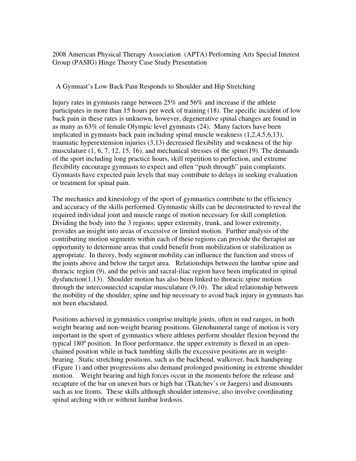
SLIDE 10 Table 1. Gymnast data and outcomes
Pain Worst : Best Oswestry16 Roland Morris17 Hip Extension ROM Left Right Shoulder Flexion ROM Left Right Shoulder External Rotation ROM Left Right Shoulder Internal Rotation ROM Left Right Initial Evaluation
8/10 : 2/10 53.33% 8/24 Hyper 3° 7° 141° 144° 51° 54° 39° 41°
8th Session (32 Days)
6° 161° 160° 68° 71° 50° 52°
Discharge (92 Days)
1/10 : 0/10 86.66% 1/24 (87.5% Improvement) 23° 19° 183° 181° 78° 78° 59° 59°