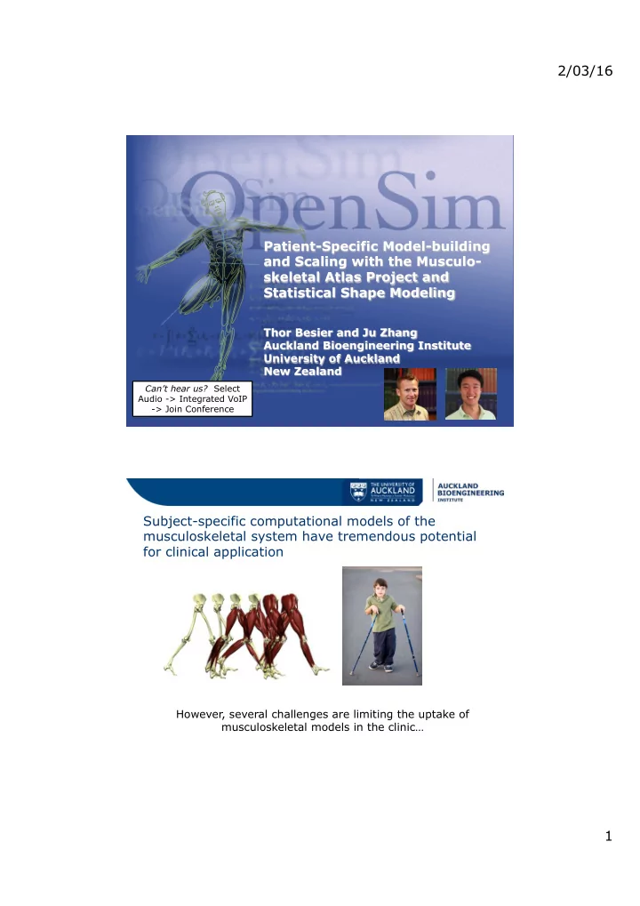

2/03/16 Patient-Specific Model-building and Scaling with the Musculo- skeletal Atlas Project and Statistical Shape Modeling Thor Besier and Ju Zhang Auckland Bioengineering Institute University of Auckland New Zealand Can’t hear us? Select Audio -> Integrated VoIP -> Join Conference Subject-specific computational models of the musculoskeletal system have tremendous potential for clinical application However, several challenges are limiting the uptake of musculoskeletal models in the clinic… 1
2/03/16 Challenges to clinical implementation Generating subject-specific models is time-consuming and costly, and requires a high level of expertise What do we mean when we say subject-specific? OpenSim – rigid body modelling Continuum mechanics – finite element modelling This talk will focus on building subject-specific bone geometry to best-match sparse motion capture and imaging data 2
2/03/16 An example problem What are the hip contact pressures during walking for this subject? + Motion capture data (mocap) MR images of the hip We want to scale or generate an OpenSim model to best- match mocap and imaging data Current approach to this problem Experimental markers Scale existing osim model Perform IK, ID, and Estimate hip contact from motion capture using anatomical CEINMS toolbox to forces using ( mocap data ) landmarks and/or estimate kinematics, MuscleForceDirection functional joint centres kinetics and muscle forces plugin s ’ C B d n a l s e e d c o r m o f E n F g i o s t s A Segment MRI of Fit mesh to point Create finite element Solve contact model pelvis cloud mesh and import into in FEBio FEBio 3
2/03/16 A different approach… Experimental markers Find set of bones that from motion capture best-match mocap AND ( mocap data ) segmented point cloud Population model of lower limb bones (n>100’s) Segment MRI of Register segmented pelvis point cloud to mocap data Overview • The MAP framework and the MAP Client • Introduction to shape modelling • Constrained scaling using shape modelling – Example 1 – scaling the hip joint with mocap – Example 2 – scaling lower limb with mocap and imaging data of femur • Muscle and joint parameters • Limitations and points for discussion • Community engagement 4
2/03/16 Our aim is to provide the biomechanics community with a tool to rapidly generate subject-specific musculoskeletal models for computational modelling The Musculoskeletal Atlas Project 5
2/03/16 Current Scaling Methods • Deform generic model to fit to landmarks • Linear (OpenSim) – Reference geometry: Delp (1990) • Linear + Nonlinear e.g. Radial Basis Functions (Anybody) – Reference geometry: Klein Horsman (2007) [Fernandez et al. 2004] [Lund et al 2015] Statistical shape models • Efficiently capture variation in shape across a population (n>100’s) = + + + PC1 PC2 PC3 Segmented population Mean of femur bones 6
2/03/16 Demo 1 – scaling the hip joint using motion capture data Results and summary of example 1 • Shape model constrains scaling to provide accurate estimate of pelvis shape and hip joint centre Registra*on ¡to ¡CT ¡image ¡ 10 ¡ Predic*on ¡Error ¡(mm) ¡ 8 ¡ 6 ¡ Linscale. ¡ 4 ¡ PC ¡Reg ¡ 2 ¡ 0 ¡ L ¡ ¡R ¡ 7
2/03/16 Example 2 – scaling the lower limb with mocap and imaging data Articulated Shape Model Degrees of freedom • Pelvis Rigid: 6 • Hip rotations: 3 • Knee flexion & abduction: 2 • Shape model scores: n Results and summary of example 2 • Shape model constrains scaling of entire lower limb to ensure an anatomically feasible solution mm Shape Iso-scaling Shape Iso-scaling Shape Iso-scaling Shape Iso-scaling model model model model 8
2/03/16 Results and summary of example 2 • Combination of marker and imaging data improves the estimation of bone geometry • Resulting bone geometry can be exported to OpenSim and/or FE packages Error Metric ¡ Proximal Partial Distal Partial Surface ¡ Surface ¡ Surface (mm) ¡ 1.75 ±0.31 ¡ 4.95 ±3.09 ¡ X (deg.) ¡ 0.15 ±0.09 ¡ 0.20 ±0.16 ¡ Y (deg.) ¡ 0.07 ±0.04 ¡ 0.06 ±0.05 ¡ Z (deg.) ¡ 0.15 ±0.08 ¡ 0.21 ±0.16 ¡ What about the muscles? • Muscle attachment sites embedded onto bones, but via points and wrapping surfaces need to be adjusted 9
2/03/16 Points for discussion • Complex joints (custom mobilizers) • Scaling muscle-tendon parameters • Body segment parameters (mass, CoM, moments of inertia) • Where are the feet and other body parts? How can you contribute? • Download the MAP Client and start developing your own plug-ins – Free and open source (GPL3 license) – Developed in Python – Cross platform https://github.com/MusculoskeletalAtlasProject/mapclient • Collaborate with us to grow our model repository (e.g. send us segmented data) • Develop plug-ins • New joint models • … 10
2/03/16 Acknowledgements • We are grateful to the Victorian Institute of Forensic Medicine (VIFM), and the Melbourne Femur Collection for providing the CT images for our shape models – John Clement – David Thomas • Auckland Bioengineering Institute – Poul Nielsen – Duane Malcolm • This work was funded by the US FDA (HHSF22320 1310119C) and NZ Ministry of Business Innovation & Employment (MBIE UOAX1407) 11
Recommend
More recommend