
10/7/16 1
Molecular and Cellular Biology
- 08. Cell Signalling :
Neurotransmitters & Receptors
- Prof. Dr. Klaus Heese
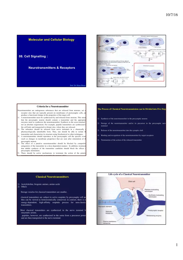
Gap junction channels, low resistance pathways between adjacent cells; direct communication between the cytoplasm of both cells, not through extracellular space (equivalent electrical circuit) (equivalent electrical circuit)
Criteria for a Neurotransmitter
Neurotransmitter are endogenous substances that are released from neurons, act on receptor sites that are typically present on membranes of postsynaptic cells, and produce a functional change in the properties of the target cell: 1) A neurotransmitter must be synthesized by and released from neurons. This means that the presynaptic neuron should contain a transmitter and the appropriate enzymes need to synthesize the neurotransmitter. Synthesis in the axon terminal is not an absolute requirement. For example, peptide transmitters are synthesized in the cell body and transported to distant sites, where they are released. 2) The substance should be released from nerve terminals in a chemically or pharmacologically identifiable form. Thus, one should be able to isolate the transmitter and characterize its structure using biochemical or other techniques. 3) A neurotransmitter should reproduce at the postsynaptic cell the specific events (such as changes in membrane properties) that are seen after stimulation of the presynaptic neuron. 4) The effect of a putative neurotransmitter should be blocked by competitive antagonists of the transmitter in a dose-dependent manner. In addition, treatments that inhibit synthesis of the transmitter candidate should block the effects of presynaptic stimulation. 5) There should be active mechanisms to terminate the action of the putative neurotransmitter (enzymatic or reuptake by neuron / glia).
The Process of Chemical Neurotransmission can be Divided into Five Steps
1) Synthesis of the neurotransmitter in the presynaptic neuron 2) Storage of the neurotransmitter and/or its precursor in the presynaptic nerve terminal 3) Release of the neurotransmitter into the synaptic cleft 4) Binding and recognition of the neurotransmitter by target receptors 5) Termination of the action of the released transmitter