
SLIDE 1
1
Enzyme-Linked-Immuno-Sorbent-Assay (ELISA)
Andre Kunert Erasmus MC - Cancer Institute Department of Medical Oncology Laboratory of Tumor Immunology Biomedical Research Techniques - November, 9th 2017
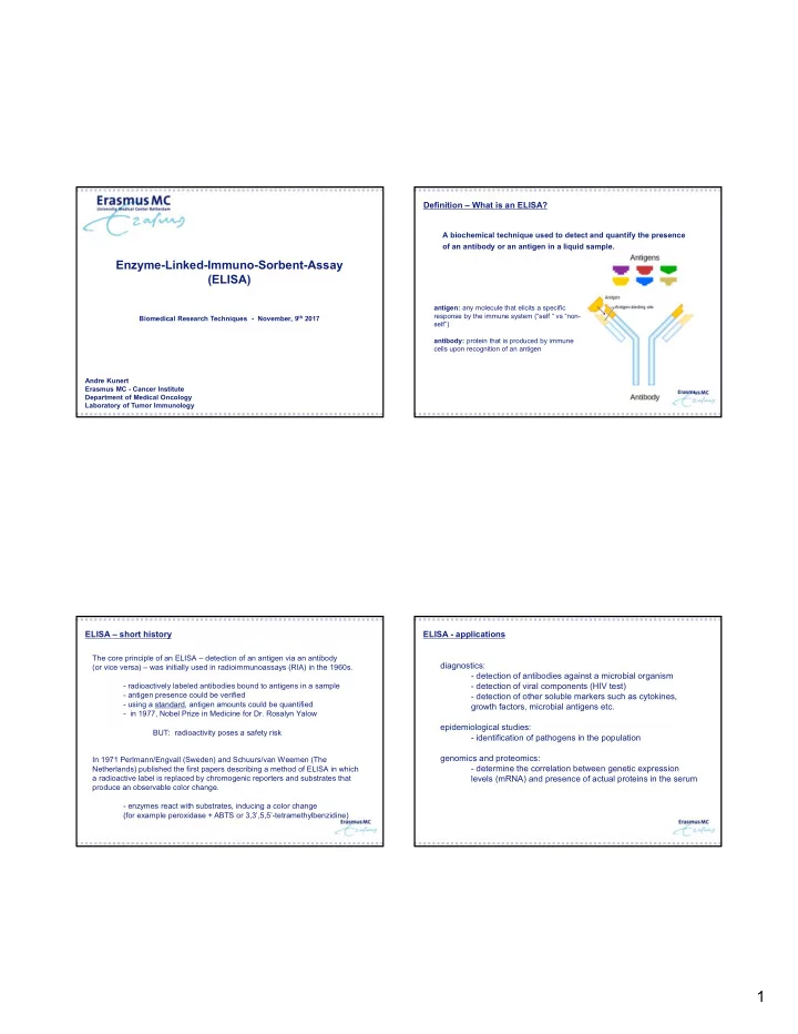
A biochemical technique used to detect and quantify the presence
- f an antibody or an antigen in a liquid sample.
Definition – What is an ELISA?
antigen: any molecule that elicits a specific response by the immune system (“self “ vs “non- self”) antibody: protein that is produced by immune cells upon recognition of an antigen
ELISA – short history
BUT: radioactivity poses a safety risk The core principle of an ELISA – detection of an antigen via an antibody (or vice versa) – was initially used in radioimmunoassays (RIA) in the 1960s.
- radioactively labeled antibodies bound to antigens in a sample
- antigen presence could be verified
- using a standard, antigen amounts could be quantified
- in 1977, Nobel Prize in Medicine for Dr. Rosalyn Yalow
In 1971 Perlmann/Engvall (Sweden) and Schuurs/van Weemen (The Netherlands) published the first papers describing a method of ELISA in which a radioactive label is replaced by chromogenic reporters and substrates that produce an observable color change.
- enzymes react with substrates, inducing a color change
(for example peroxidase + ABTS or 3,3’,5,5’-tetramethylbenzidine)
ELISA - applications diagnostics:
- detection of antibodies against a microbial organism
- detection of viral components (HIV test)
- detection of other soluble markers such as cytokines,
growth factors, microbial antigens etc. epidemiological studies:
- identification of pathogens in the population
genomics and proteomics:
- determine the correlation between genetic expression