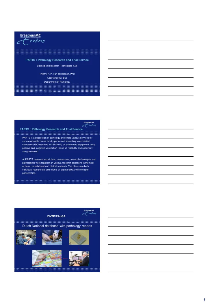

PARTS : Pathology Research and Trial Service Biomedical Research Techniques XVII Thierry P. P. van den Bosch, PhD Kadir Akdeniz, BSc Department of Pathology PARTS : Pathology Research and Trial Service PARTS is a subsection of pathology and offers various services for very reasonable prices mostly performed according to accredited standards (ISO-standard 15189:2012) on automated equipment using positive and negative verification tissue so reliability and specificity are guaranteed. At PARTS research technicians, researchers, molecular biologists and pathologists work together on various research questions in the field of basic, translational and clinical research. The clients are both individual researchers and clients of large projects with multiple partnerships. DNTP/PALGA Dutch National database with pathology reports 1
Public database PODB (Public Pathology Database) PALGA DNTP Dutch National Tissuebank Portal Online portal for request and delivery of pathology data and samples How to request pathology samples? https://aanvraag.palga.nl Request form: Attachment containing Pathology numbers 2
Any questions regarding Pathology data / samples? dntp@erasmusmc.nl Fixation and processing of tissues Good standardized tissue preservation is very important for further research. We fixate tissue in formalin for 24 hours and they are processed using automated tissue processor overnight. The tissue processor processes the material in a number of steps, including an after- fixation in formalin, an ascending alcohol sequence, which removes the water and xylene treatment that removes the ethanol and makes the tissue receptive to the paraffin. Gross-room: fresh tissue preparation Imbedding of processed tissue Histology - Histochemistry In the PARTS laboratory technicians work who are very skilled at cutting sections. Their experience allows them to process paraffin blocks quickly and with high quality into sections that can be used for many purposes The histology laboratory is equipped with iso-certified equipment with which not only the standard hematoxylin & eosin (H&E) section can be made, but also various special stainings such as PAS or Sirius Red. Additional blank sections can be made if it is not yet known which staining or analyses will follow. For DNA, RNA or protein isolation sections are collected in Eppendorf tubes. If laser microdissection is desired, the sections are prepared on membrane slides. 4 µm tissue sections HE slides with corresponding tissue blocks PAS staining 3
Immunohistochemistry In addition to histochemical staining, immunohistochemical (IHC) staining and immunofluorescence (IF) are also possible on tissue sections. For IHC and IF we make use of the automatic color stations Ventana BenchMark Ultra and the Ventana Discovery. These platforms have been developed for the fully automatic execution of IHC and IF on histological or cytological samples. PARTS works with validated antibodies. When using non-routinely used antibodies and antibodies that have recently been developed, they must be validated first. Ventana Ultra benchmark Pan-keratine staining GFAP staining Multiplex Immunofluorescence With the multiplex IF method it is possible to detect two proteins in one section. With multiplex-IF it is currently possible to co-localize a maximum of four proteins, each protein being given its own label. This panel stained tonsil tissue with CD8 (DCC- blue), CD4 (Red610- red), CD68KP1 (Cy5- white) and CD20 (FAM, green) Erasmus MC Tissue Bank Hospital Intergrated Biorepository, integral part of the Clinical Pathology Department 100.000 Frozen tissue (left-over / residual) - Surgical specimen - Biopsies (routine) - Post Mortem 3 million FFPE archival material Request for frozen/fresh/FFPE tissue contact m.oomen@erasmusmc.nl or p.riegman@erasmusmc.nl. 4
Molecular pathology Techniques and support for molecular analyzes of human or animal material operational within the laboratory: DNA/RNA isolation * from among others: FFPE, frozen tissue, routine (stained) sections, blood, serum, plasma, urine and cell cultures * manual microdissection of tissue sections ABI sequencer * Sanger sequencing * Mutation specific PCR * SNaPshot analyses * Fragment analyses * MSI analyses * Tissue / cell line identification (STR genotyping) Next generation sequencing (Ion Torrent) * Targeted analyses with custom made panels of DNA, RNA and liquid biopsies * Bioinformatics support for generating and analysis of data In situ hybridisatie * FISH, CISH * mRNA expressie (RNAscope) * microRNA expressie (Ventana Benchmark Discovery) microRNA ISH U6 Virtuel microscopy A virtual microscope can digitally store images of tissue sections with a high resolution. The Pathology department has a Hamamatsu Nanozoomer 2.0 HT digital slide scanner(virtual microscope)for scientific use. The NDP viewer software allows you to view the images in all enlargements and to export parts of them for publication. Measurements and annotations can also be made. With additional image processing software it is possible to further analyze images. You can also make the images available and have them assessed by others via intranet within the Erasmus MC or via the internet. After a short course by one of our PARTS employees, you can use the Nanozoomer virtual microscope yourself. It is also possible that PARTS employees scan your slides. Hamamatsu Nanozoomer Visiomorph digital analysis software Tissue Micro Arrays High-throughput molecular analysis of tissues is possible with the tissue microarray (TMA) technology A TMA contains hundreds of tissue pieces from original donor paraffin blocks. With 1 section of a TMA you analyze all tissues simultaneously with a small amount of reagent. TMAs are suitable for morphological studies, histochemistry, immunohistochemistry, and DNA and RNA in situ hybridization. PARTS has a fully automatic TMA Grand Master. Researchers can, after an introductory course, make their own TMAs. PARTS employees can do this work for you. The TMA grand master also offers the possibility to capture the cores in Eppendorf tubes for other molecular research Manual TMA machine Fully automated TMA Grandmaster TMA blocks 5
Laser Capture Microdissection The PALM laser microdissection technique can isolate single cells or histologically pure cell populations from tissue sections that are important for your research. The cells of interest can be separated from the rest by means of microdissection and used for, among other things, DNA genotyping, RNA analysis, cDNA generation and proteomics. Microdissection is possible on both FFPE and fresh frozen tissue with normal or fluorescence light. The dissection is possible from existing sections. If the section is to be made, it is best to use specially made membrane slides. After an introductory course, you can use the equipment on your own. PALM Electron microscopy Electron microscopy is a very powerful technique to study the internal structure or the surface morphology of materials and biological objects. Electron microscopy makes it possible to observe details of a few nanometers. PARTS provides electron microscopy techniques with a focus on sample preparation and image analysis. PARTS has a Morgagni 268 D, Transmission Electron Microscope to observe thin sections. PARTS can help you to determine optimal experimental conditions for your project. We have experience with a wide range of biological specimens. Techniques: * Chemical fixation * Plastic embedding * Ultramicrotomy * Electron microscopy imaging * Image analysis Pathology Research Trial Service Clinical Trial: For research on material for the purposes of a clinical trial is the concent of the patient in the form of an Informed Concent needed and approval of the METC , both is the responsibility of the Trial coordinator. The results of the at the pathology outsourced research may directly affect the subsequent treatment of the patient in the trial and will have to be carried out with a certain urgency. Before a trial officially can be started, the responsible and competent research technician will review the adoption of the trials. Contact Trials: Monique Oomen m.oomen@erasmus.nl Frontoffice PARTS parts@erasmusmc.nl 6
Contact information Website: https://intranet.erasmusmc.nl/pathologie/partsinfo/ Clinical Trials: PARTS@erasmusmc.nl Presenter contact: t.vandenbosch@erasmusmc.nl 7
Recommend
More recommend