Unusual presentation of complete tracheal rings in a 15 year old - - PDF document

unusual presentation of complete tracheal rings in a 15
SMART_READER_LITE
LIVE PREVIEW

Unusual presentation of complete tracheal rings in a 15 year old - - PDF document

Pediatric Anesthesia and Critical Care Journal 2015; 3(2):124-128 doi:10.14587/paccj.2015.25 Unusual presentation of complete tracheal rings in a 15 year old trauma patient B. S. Schmidt 1 , E. J. Herschmiller 2 , R. J. Jarrah 1 , T. A. Nakagawa


slide-1
SLIDE 1

Pediatric Anesthesia and Critical Care Journal 2015; 3(2):124-128 doi:10.14587/paccj.2015.25 Schmidt et al. Complete tracheal rings

124

Key points Complete tracheal rings may be encountered in the spectrum of congenital tracheal stenosis and is usually diag- nosed in early childhood. In rare cases they can remain undiagnosed throughout childhood and manifest as an unexpectedly difficult airway requiring intubation with a much smaller endotracheal tube than would be expected for the patient’s size and age

Unusual presentation of complete tracheal rings in a 15 year old trauma patient

  • B. S. Schmidt1, E. J. Herschmiller2, R. J. Jarrah1, T. A. Nakagawa1

1Department of Anesthesiology (Section on Pediatric Critical Care) and Surgery, Wake Forest School of Medicine, Win-

ston-Salem, North Carolina, USA

2Department of Anesthesiology, New York Presbyterian/Columbia, New York, USA

Corresponding author: 1T. A. Nakagawa, Department of Anesthesiology (Section on Pediatric Critical Care) and Surgery, Wake Forest School of Medicine, Winston-Salem, North Carolina, USA. Email: tnakagaw@wakehealth.edu Abstract Complete tracheal rings may be encountered in the spec- trum of congenital tracheal stenosis, and is usually dia- gnosed in early childhood. We present an unusual case

  • f a 15-year-old trauma patient with progressive respira-

tory failure and an unanticipated difficult airway during

  • intubation. The patient progressed to cardiopulmonary

arrest and required extracorporeal membrane oxygena- tion for respiratory support. Bronchoscopy revealed complete tracheal rings impeding passage of an appro- priately sized endotracheal tube into the airway. A much smaller endotracheal tube was required to intubate this patient’s trachea. Keywords: Intubation, intratracheal, tracheal stenosis, extracorporeal membrane oxygenation, bronchoscopy, tracheal abnormalities. Introduction Complete tracheal rings are a finding that may be en- countered in the spectrum of congenital tracheal steno-

  • sis. Disproportionate growth of the tracheal cartilage

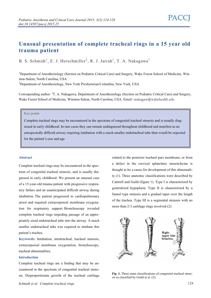
related to the posterior tracheal pars membrane, or from a defect in the cervical splanchnic mesenchyme is thought to be a cause for development of this abnormali- ty (1). Three anatomic classifications were described by Cantrell and Guild (figure 1). Type I is characterized by generalized hypoplasia. Type II is characterized by a funnel type stenosis and a gradual taper over the length

  • f the trachea. Type III is a segmental stenosis with no

more than 2-3 cartilage rings involved (2).

  • Fig. 1. Three main classifications of congenital tracheal steno-

sis as classified by Guild et al. (2).

slide-2
SLIDE 2

Pediatric Anesthesia and Critical Care Journal 2015; 3(2):124-128 doi:10.14587/paccj.2015.25 Schmidt et al. Complete tracheal rings

125

Other types of tracheal stenosis have been described, including a “corkscrew” type of stenosis of the distal trachea (3). Concentric tracheal rings are a common characteristic of each of the many types of congenital tracheal stenosis. An abnormal origin of the right upper lobe bronchus arising directly from the trachea (bron- chus suis or “pig bronchus”) is seen in up to 20% of ca- ses. We present an unusual case of a 15-year-old trauma pa- tient with progressive respiratory failure and an unanti- cipated difficult airway during intubation due to undia- gnosed complete tracheal rings. We received parental permission to publish this case report. Further, the Wake Forest Institutional Review Board waives the need for consent for case reports provided they comply with HIPAA regulations. Case report A 15 year old male with a history of hypothyroidism and Scheuermann’s kyphosis presented to our pediatric trauma center after suffering an all-terrain vehicle acci-

  • dent. Injuries included: three-column fracture of his spi-

ne with cord transection at the level of T8-T9, pulmona- ry contusions, and rib fractures adjacent to the spinal

  • fractures. The patient was admitted to the pediatric in-

tensive care unit (PICU) for blood pressure management and neurologic monitoring, with no respiratory com-

  • promise. He was taken to the operating room for lami-

nectomy, decompression of the spinal cord, and poste- rior spinal fusion. He was easily ventilated using a bag mask during induction. The patient’s cervical collar was removed, and inline cervical stabilization was maintai- ned while an asleep elective fiberoptic intubation was

  • performed. A 7.5 cuffed endotracheal tube (ETT) was

placed without difficulty, and secured at 23 cm. Endo- tracheal tube position was confirmed by auscultation and continuous end-tidal carbon dioxide (ETCO2) moni-

  • toring. He was placed in the prone position for the po-

sterior spinal fusion. Postoperatively, he was extubated and returned to the PICU with no cardiorespiratory is-

  • sues. On postoperative day 3 he developed dyspnea with

acute rapidly progressive hypoxia. Oxygen saturations decreased to the 70s, and did not improve despite ag- gressive pulmonary toilet and institution of high-flow nasal cannula oxygen therapy. Due to persistent hy- poxemia and impending respiratory failure, rapid se- quence induction was performed with etomidate and succinylcholine to electively secure his airway. The ini- tial intubation attempt was unsuccessful; the provider was able to pass a 7.5-mm ETT through the glottic ope- ning; however, more distal (subglottic) resistance resul- ted in herniation of the ETT back into the laryngeal ve-

  • stibule. A second intubation attempt was successful with

placement of the 7.5 cuffed ETT in the airway confir- med with a colorimetric ETCO2 detector. However,

  • xygenation saturations failed to improve. Direct la-

ryngoscopy was performed to evaluate ETT position. This examination revealed a Grade 2 view of the ETT passing under the epiglottis and through the vocal cords. Because of persistent desaturation, the ETT was remo- ved, and bag-mask-ventilation was reinitiated, with mild improvement in oxygen saturations. Reintubation with a 7.5 cuffed ETT resulted in color change on the ETCO2 detector when ventilation was initiated. However, despi- te aggressive manual ventilation with 100% oxygen, sa- turations did not improve and the patient suffered a bradycardic arrest. Pediatric Advanced Life Support (PALS) measures were initiated. Bilateral breath sounds were minimally audible with intermittent oxygen satura- tions in the 50s. Video laryngoscopy was performed with a McGrath size 4 laryngoscope to confirm position of the ETT due to the difficult intubation. The ETT tip was visualized sit- ting outside the glottic opening in the laryngeal vestibu-

  • le. The ETT was removed and bag mask ventilation re-
  • sumed. Repeat direct laryngoscopy (DL) with the same

laryngoscope provided a Grade 1 view and a 7.0 cuffed ETT was visualized passing through the vocal cords. Oxygen saturations improved to the mid-60s with ma- nual ventilation and return of spontaneous circulation

slide-3
SLIDE 3

Pediatric Anesthesia and Critical Care Journal 2015; 3(2):124-128 doi:10.14587/paccj.2015.25 Schmidt et al. Complete tracheal rings

126

(ROSC) occurred after 7 min of cardiopulmonary resu- scitation. Chest radiograph revealed opacification of the entire left chest, concerning for hemothorax, and a chest tube was placed with evacuation of 800 ml of bloody output. This did not appreciably improve saturations and a flexible bronchoscopy was performed to evaluate for mucus plugs or kinking of the ETT. Despite poor visualization

  • f the airway, it appeared that the ETT was unobstruc-
  • ted. Persistent hypoxia despite aggressive airway ma-

neuvers resulted in a decision to pursue extracorporeal membrane oxygenation (ECMO) support. Percutaneous cannulation of the right internal jugular vein and right common femoral vein was performed without complica-

  • tion. Veno-venous (VV) ECMO support was initiated

and oxygen saturations quickly improved. Further attempts to advance the ETT were unsuccessful. Flexible bronchoscopic evaluation was repeated sho- wing the ETT positioned well above the carina (figure 2). Complete tracheal rings were found to comprise the lower two-thirds of the airway (figure 3). The tracheal rings caused a long segment of tracheal stenosis which impeded further advancement of the ETT.

  • Fig. 2. Tip of 7.5 French endotracheal tube at point of maxi-

mal advancement, well above the carina.

  • Fig. 3. Concentric tracheal rings observed more clearly after

removal of the endotracheal tube while fully supported on ex- tracorporeal membrane oxygenation.

  • Fig. 4. Three-dimensional reconstruction of the patient’s upper

airway and proximal tracheal from computed-tomographic images, demonstrating funnel-like narrowing of trachea with a subtle appearance of concentric cartilaginous rings.

A 5.0 uncuffed ETT was placed under direct broncho- scopic visualization and advanced 1-2 cm above the ca-

  • rina. No air leak was noted when this smaller tube was

placed in the airway. Review of the initial computed tomography (CT) scan revealed subtle evidence of com- plete tracheal rings in the distal trachea. These findings were more noticeable after advanced three-dimensional reconstructions were created (figure 4). Repeat chest CT scan did not reveal any intra- or extrathoracic causes for the sudden decompensation or the hemothorax. Cardiac work-up did not reveal any congenital anomalies, ab-

slide-4
SLIDE 4

Pediatric Anesthesia and Critical Care Journal 2015; 3(2):124-128 doi:10.14587/paccj.2015.25 Schmidt et al. Complete tracheal rings

127

normal ventricular function, or pulmonary arterial sling. The patient was weaned from VV-ECMO to minimal mechanical ventilator support over the ensuing two days. Unfortunately, neurologic examination revealed that the patient was in a persistent vegetative state and unre- sponsive despite weaning sedation. Head imaging revea- led evidence of global anoxic injury, and a grim progno- sis for meaningful neurologic recovery was conveyed to his family. After further discussion, supportive medical therapies were terminated and the patient expired on hospital day

  • 11. Request for autopsy was declined.

Discussion Congenital tracheal stenosis is frequently a diagnosis of

  • infancy. The hallmark symptom is biphasic stridor

which can be exacerbated by an upper respiratory infec-

  • tion. Diagnosis can also be made at the time of opera-

tion for an unrelated issue, as intubation can be difficult (4) or even impossible, requiring the use of laryngeal mask airway or advanced invasive support such as

  • ECMO. Typically, complete tracheal rings are diagno-

sed and monitored over time by bronchoscopy. Boiselle et al. suggest that paired end-inspiratory dynamic expi- ratory CT techniques with 3-D reconstruction may pro- vide just as much information as bronchoscopy and can be used as an adjunct (5). In young children, however, dynamic expiratory CT techniques may not be reliable since they require patient cooperation and compliance with breathing instructions. Definitive management of complete tracheal rings will require surgical intervention in the majority of patients. Slide tracheoplasty is the treatment of choice (6). When an open approach is not safe or advisable, endoscopic approaches may be consi-

  • dered. Laser division allows a controlled separation of

the complete rings along the posterior wall of the tra- chea (7). Balloon dilatation under fluoroscopy has been used to successfully divide the posterior aspect of the complete cartilaginous rings (8). Both techniques often involve the postoperative placement of a tracheal stent, and usually require repeat interventions to achieve a suf- ficiently wide airway (9). Pediatric cardiothoracic surge- ry and ECMO support should be available in the event

  • f airway compromise.

Tracheal rings are associated with vascular anomalies, most commonly a pulmonary sling (9). Other anomalies have been reported including tracheoesophageal fistula, esophageal atresia, VATER/VACTERL syndromes, cardiac abnormalities, Pfeiffer’s syndrome (10), and scoliosis (1). Our case is somewhat unique since this patient was asymptomatic during childhood. This child never de- monstrated stridor and was able to maintain an active lifestyle without breathing problems or evidence of air- way obstruction. Importantly, our case illustrates a rare cause of an unexpected difficult airway. Prior documen- tation of intubation did not suggest a difficult airway; however, fiberoptic intubation was used to provide cer- vical spine protection. There was nothing to suggest tra- cheal stenosis during previous airway endoscopy. No problems with increased airway pressures, oxygenation,

  • r ventilation were documented in the intraoperative

anesthesia record. We suspect intubation during the first surgery appeared unremarkable because the 7.5 cuffed ETT with the balloon inflated compressed the vocal cords, preventing movement of the ETT, which was likely positioned at the level of the thoracic inlet. References

  • 1. Li Y, Khambatta HJ, Stone JG, Mets B. Unsuspec-

ted concentric tracheal rings in a 14-year-old with

  • scoliosis. Br J Anaesth. 2002; 88:732-4.
  • 2. Cantrell JR, Guild HG. Congenital Stenosis of the
  • Trachea. Am J Surg. 1964; 108:297-305.
  • 3. Bryant R III, Morales DL. Corkscrew trachea: a

novel type of congenital tracheal stenosis. Ann Thorac Surg. 2009; 87:1923-5.

  • 4. Trivedi P, Hardy C. A case of an unexpected airway

difficulty in the cardiac

  • perating

room. CCAS E-News. The Congenital Cardiac Anesthesia Society. Summer 2011. Available at:

slide-5
SLIDE 5

Pediatric Anesthesia and Critical Care Journal 2015; 3(2):124-128 doi:10.14587/paccj.2015.25 Schmidt et al. Complete tracheal rings

128

http://www.ccasociety.org/newsletters/2011summer /case.html. Accessed November 13, 2014.

  • 5. Boiselle PM, Ernst A, DeCamp MM. CT diagnosis
  • f complete tracheal rings in an adult. J Thorac
  • Imaging. 2007; 22:169-71.
  • 6. Terada M, Hotoda K, Toma M, Hirobe S, Kamaga-

ta S. Surgical management of congenital tracheal

  • stenosis. Gen Thorac Cardiovasc Surg. 2009;

57:175-83.

  • 7. Blackmore K, Kubba H, Clement WA. Laser divi-

sion of congenital complete tracheal rings. Int J Pe- diatr Otorhinolaryngol. 2010; 74:1327-30.

  • 8. Jaffe RB. Balloon dilation of congenital and acqui-

red stenosis of the trachea and bronchi. Radiology. 1997; 203:405-9.

  • 9. Ho AS, Koltai PJ. Pediatric tracheal stenosis. Otola-

ryngol Clin North Am. 2008; 41:999-1021.

  • 10. Faust RA, Stroh B, Rimell F. The near complete

tracheal ring deformity. Int J Pediatr Otorhinola-

  • ryngol. 1998; 45:171-6.