Tricuspid Regurgitation: No Disclosures Implication for Left-sided - - PDF document

tricuspid regurgitation
SMART_READER_LITE
LIVE PREVIEW

Tricuspid Regurgitation: No Disclosures Implication for Left-sided - - PDF document

Tricuspid Regurgitation: No Disclosures Implication for Left-sided Procedures Chari Y.T. Hart MD, FACC Interventional Echocardiography Queens Heart Physician Practice Queens Medical Center-Honolulu, Hawaii Assistant Clinical Professor


slide-1
SLIDE 1

1

No Disclosures

Tricuspid Regurgitation:

Implication for Left-sided Procedures

Chari Y.T. Hart MD, FACC

Interventional Echocardiography

Queen’s Heart Physician Practice Queen’s Medical Center-Honolulu, Hawaii Assistant Clinical Professor of Medicine, John A. Burns School of Medicine, University of Hawaii

October 01, 2016

Innovative Procedures, Devices, and State of the Art Care for Arrhythmias, Heart Failure and Structural Heart Disease
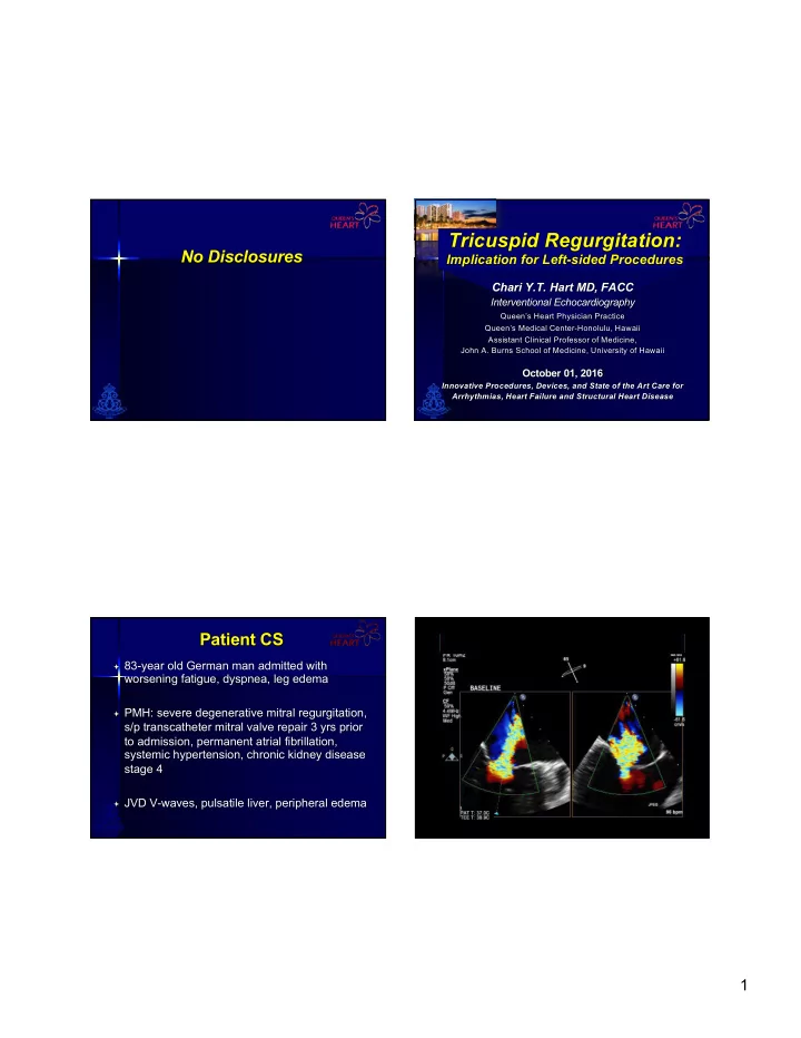
Patient CS

ª 83-year old German man admitted with

worsening fatigue, dyspnea, leg edema

ª PMH: severe degenerative mitral regurgitation,

s/p transcatheter mitral valve repair 3 yrs prior to admission, permanent atrial fibrillation, systemic hypertension, chronic kidney disease stage 4

ª JVD V-waves, pulsatile liver, peripheral edema

slide-2
SLIDE 2

2

slide-3
SLIDE 3

3

Apical 2-chamber view MitraClip, mild residual MR Mitral inflow diastolic MG 4 mmHg at 61 bpm Parasternal LAX view Preserved LVEF, MitraClip, mild MR Apical 4-chamber view dilated RV, RA; severe TR Subcostal view of IVC, hepatic vein systolic flow reversals RV tissue Doppler peak systolic velocity Hepatic vein PW Doppler systolic flow reversals

The Clinical Problem…

ª Incidence of tricuspid regurgitation

associated with left-sided valvular disease 8% to 35% of cases

ªMore common in mitral valve disease ªAlso in aortic valve disease

ª Tricuspid regurgitation is often clinically

silent, thus an “ignored” problem in left- sided disease

Jacovella G et al. Cardiol Prat 1971;22:235-41 King RM et al. Circulation 1984;70:1193-7

Outline

² Anatomy of the tricuspid valve ² Primary versus secondary (functional)

tricuspid regurgitation (TR)

² Pathophysiology of secondary tricuspid

regurgitation

² Prognosis of tricuspid regurgitation ² Guideline indications for intervention of

secondary tricuspid regurgitation

² Future non-surgical options for tricuspid

regurgitation

slide-4
SLIDE 4

4

n 3 leaflets (septal,

anterior, posterior)

n The annulus n The chordae n The papillary

muscles

n The adjacent

myocardium (atrial, ventricular)

Tricuspid Valve Anatomy

Dreyfus G.D., Corbi PJ, Chan KM, et al. Ann Thorac Surg 2005;79:127-32

n Largest valve n Area 4-6 cm2 n Diam 27-29mm n Inferior, anterior to

the MV

n Saddle-shaped

ellipsoid becomes planar & circular as it dilates (A-P)

Dreyfus G.D., Corbi PJ, Chan KM, et al. Ann Thorac Surg 2005;79:127-32

Tricuspid Valve Anatomy Tricuspid Regurgitation

Primary 20%

Secondary “Functional” 80%

Rheumatic Degenerative (myxomatous) Congenital (Ebstein’s, dysplasia, cleft, etc.) Infective Endocarditis Radiation Carcinoid Blunt chest trauma Iatrogenic trauma:

  • RV biopsy
  • RV PM or ICD leads

Left heart disease

  • Left ventricular dysfunction
  • Mitral or aortic valve disease

Right ventricular dysfunction

  • RV cardiomyopathy
  • RV ischemia
  • RV volume overload (ASD,

APVR) Pulmonary Hypertension

  • Chronic lung disease
  • Left to right shunt
  • Pulmonary

thromboembolism Right atrial abnormalities

  • Atrial fibrillation

RHD

Pathophysiology of Tricuspid Regurgitation in Mitral Valve Disease

Ischemic DCM MS MR á Left atrial size á Left atrial pressure Atrial fibrillation Pulmonary hypertension Tricuspid annulus dilatation (tethering of TV) RV dilatation & dysfunction TRICUSPID REGURGITATION Organic TV disease

Shiran A, Sagie A. J Am Coll Cardiol 2009;53:401-8

slide-5
SLIDE 5

5

RHD

Pathophysiology of Tricuspid Regurgitation in Mitral Valve Disease

Ischemic DCM MS MR á Left atrial size á Left atrial pressure Atrial fibrillation Pulmonary hypertension Tricuspid annulus dilatation (tethering of TV) RV dilatation & dysfunction TRICUSPID REGURGITATION Organic TV disease

Charles J. Bruce, and Heidi M. Connolly Circulation. 2009;119:2726-2734

1.0 0.8 0.6 0.7 0.2 0.0 200 400 600 800 1,000 1,200 1,400 Surviving Tricuspid regurgitation Days None Mild Moderate Severe

Severe Tricuspid Regurgitation Significant â exercise capacity (low output) Negative impact on long-term survival

>5,000 patients

Prognostic Implications of TR in patients with VHD

ª Moderate+ secondary TR, mean age 68+16 yrs ª Mean follow-up 43 + 24 mos:

ªDeaths 13 (14%) ªSurgeries 12 (13%) ªMultivariate analysis, predictors of death:

ª RVSP (p=0.0038)

ªRVSP > 48 mmHg, associated with significantly increased mortality and decreased surgery-free survival

ª Device implantation (p=0.0487)

Jhawar MB et al. J Heart Valve Dis 2013; 418-24

What do the Guidelines Say?

J Am Coll Cardiol. 2014;63(22):2438-2488. doi:10.1016/j.jacc.2014.02.537 *TA dilation is defined by >40 mm on TTE (>21 mm/m2) or >70 mm on direct intraoperative

slide-6
SLIDE 6

6

2014 AHA/ACC Valvular Heart Disease Guidelines Intervention Recommendations for Tricuspid Regurgitation Class I (…is recommended/indicated):

ª Tricuspid valve surgery is recommended for patients with

severe functional TR (stages C and D) undergoing left- sided valve surgery.

J Am Coll Cardiol. 2014;63(22):2438-2488. doi:10.1016/j.jacc.2014.02.537

2014 AHA/ACC Valvular Heart Disease Guidelines Intervention Recommendations for Tricuspid Regurgitation Class IIa (…is reasonable/can be beneficial):

ª Tricuspid valve repair can be beneficial for patients with

mild, moderate or greater (stage B) at time of left-sided valve surgery with either 1) tricuspid annular dilatation* or 2) prior evidence of right heart failure. *Tricuspid annulus dilatation of >40mm diameter or 21 mm/m2 on

preop TTE; >70mm direct intraop

ª Tricuspid valve repair can be beneficial for patients with

symptoms due to severe primary TR that are unresponsive to medical therapy

J Am Coll Cardiol. 2014;63(22):2438-2488. doi:10.1016/j.jacc.2014.02.537

2014 AHA/ACC Valvular Heart Disease Guidelines Intervention Recommendations for Tricuspid Regurgitation Class IIb (…may be reasonable/may be beneficial):

ª Tricuspid valve repair may be considered for

asymptomatic or minimally symptomatic patients with severe primary TR (stage C) and progressive moderate or greater RV dilatation and/or systolic dysfunction.

ª Tricuspid valve repair may be beneficial for patients with

persistent symptoms due to severe TR (stage D) in patients who have undergone previous left-sided valve surgery and who do not have severe pulmonary hypertension or significant RV systolic dysfunction.

J Am Coll Cardiol. 2014;63(22):2438-2488. doi:10.1016/j.jacc.2014.02.537

Pilar Tornos Mas et al. Heart 2015;101:1840-1848

Multimodality Imaging in the Assessment of Tricuspid Regurgitation and Right Ventricular Volumes and Function

slide-7
SLIDE 7

7

Before left-sided valve surgery, careful assessment of TR severity, tricuspid annulus measurement TA diameter in 4-ch view septal-lateral dimension

Future Non-Surgical Options for Tricuspid Regurgitation

Josep Rodés-Cabau, MD, Rebecca T. Hahn, MD Azeem Latib, MD et al. J Am Coll Cardiol 2016;67:1829–45

Future Non-Surgical Options for Tricuspid Regurgitation

Josep Rodés-Cabau, MD, Rebecca T. Hahn, MD Azeem Latib, MD et al. J Am Coll Cardiol 2016;67:1829–45

Challenging Anatomy of the Tricuspid Valve for Transcatheter Therapies

n Large tricuspid annulus n Nonplanar, elliptical n Fragile TA tissue, narrow

annular shelf

n Noncalcified annulus n Angulation with SVC, IVC n Trabeculated RV, mucular

bands, chordae

n Thin RV free wall n Proximity of AV node, right

His bundle branch

n Proximity of the RCA to

annulus, risk of injury

n Risk of occlusion of

coronary sinus, vena cava,

  • utflow tract

n Slow-flow in RV n Pacemaker, ICD leads

slide-8
SLIDE 8

8

Future Non-Surgical Options for Tricuspid Regurgitation

Christoph Hammerstingl, Robert Schueler, Margarita Malasa et al. Eur Heart J. 2016 Mar 7;37(10):849-53

Interventional Edge-to-Edge Repair with the MitraClip system

n

3 patietnts wih severe, symptomatic tricuspid regurgitation

n

Feasibility, safety demonstrated

n

âERO with 3D-TEE, áLV stroke volume, âN-terminal proBNP, relief of clinical symptoms for right heart failure

n

Promising option for patients at prohibitive surgical risk

Conclusions

ª The tricuspid valve is COMPLEX ª The majority (80%) of tricuspid regurgitation is

secondary (functional)

ª Have R-E-S-P-E-C-T for tricuspid regurgitation

secondary to left heart pathology, not automatically corrected

ª Less emphasis on conservative treatment, importance

  • f concomitant correction with left heart procedures

ª Tricuspid annular dilatation is progressive, ‘prophylactic’

intervention should be considered, regardless of TR severity

ª Proactive approach to secondary tricuspid regurgitation

and its surgical/transcatheter correction

ª Improved long-term survival outcomes for patients

Ma Mahalo fo for y r your a r atten ttentio tion!

slide-9
SLIDE 9

9

Josep Rodés-Cabau, MD, Rebecca T. Hahn, MD Azeem Latib, MD et al. J Am Coll Cardiol 2016;67:1829–45

Caval Valve Implantation (CAVI) Tric Valve Self-Expandable

Josep Rodés-Cabau, MD, Rebecca T. Hahn, MD Azeem Latib, MD et al. J Am Coll Cardiol 2016;67:1829–45

Caval Valve Implantation (CAVI) Balloon Expandable Valves

Josep Rodés-Cabau, MD, Rebecca T. Hahn, MD Azeem Latib, MD et al. J Am Coll Cardiol 2016;67:1829–45

Coaptation Device FORMA Repair System

Josep Rodés-Cabau, MD, Rebecca T. Hahn, MD Azeem Latib, MD et al. J Am Coll Cardiol 2016;67:1829–45

The Mitralign Device

slide-10
SLIDE 10

10

Josep Rodés-Cabau, MD, Rebecca T. Hahn, MD Azeem Latib, MD et al. J Am Coll Cardiol 2016;67:1829–45

The TriCinch System