
SLIDE 1
The CRE Luc Reporter M ouse M odel A transgenic bioimaging mouse - - PowerPoint PPT Presentation
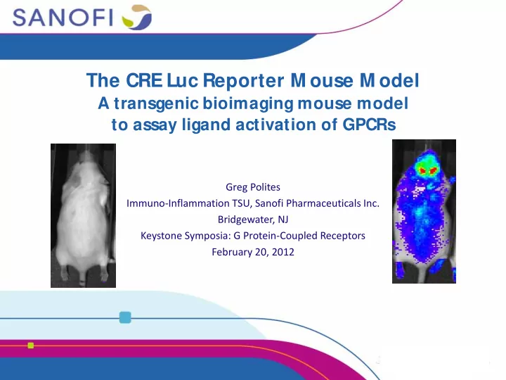
The CRE Luc Reporter M ouse M odel A transgenic bioimaging mouse model to assay ligand activation of GPCRs Greg Polites Immuno-Inflammation TSU, Sanofi Pharmaceuticals Inc. Bridgewater, NJ Keystone Symposia: G Protein-Coupled Receptors
2
Optics CCD Bioluminescent Source
IP3 Ca2+ as b g AC cAM P PKA
CREB
PLC
DAG PKC ai b g aq b g CaM K
luci
+
3
In vivo In vitro Assay Dev Target ID Development Activity of selected compound in vivo (PK, PD), tissues or whole body imaging Source of cells for in vitro assays Clinical
4
Feasibility profiles T,B,macs. Pancreas Lung CNS Specific pilots to reach project application decision No-Go Go No-Go Go No-Go Go Project application 64 11, 16
CRE-Luc Liver Kidney Adipose 187 219 229 175
5
IVIS bioimaging Whole live animal imaging Simple, quick Limited resolution
Microplate reader Sensitive, accurate Better organ resolution Time-consuming
6
brain
baseline isoproterenol 100000 200000 300000 400000 500000
15X ***
treatment p/s/cm2
spinal cord
baseline isoproterenol 50000 100000 150000 200000
15X ***
treatment p/s/cm2
ROI
7
CH1_DATA(cpm) 2000 4000 6000 8000 10000 12000 14000 16000 18000 20000
1440 2880 4320 5760 7200 8640 10080 11520 12960 14400
CH1_DATA(cpm) 6000 7000 8000 9000 10000 11000 12000 13000 14000 15000 16000
1440 2880 4320 5760 7200 8640 10080 11520 12960 14400
1.93x
8
DMSO 10uM 1000 2000 3000 4000 * * * p<0.0001 * * * 11X [isoproterenol] cps
media 3uM 10uM 50 100 150 200 250 300 350 400 450 500 550 * * 3X * * p<0.01 [dopamine] cps
n e u r on s
10uM F media DMSO F 1nM 10nM 100nM 1uM 5000 10000 15000 20000 25000 30000 35000 ns ns 40% 38% * * * * 9X
* * p<0.005
[AMN082] cps
DMSO F/R 100nM 1uM 10uM 50000 100000 150000 45%↓ 39%↓ 20%↓
* * * p<0.0005 * * p<0.005 * p<0.05
10uM F/ 10uM R 30X [CP55940] cps
9
100 200 300 400 500 600 700 2 4
Days Glucose (mg/ dL)
Control STZ
STZ increases blood glucose STZ (day 4) blocks the induction of luci by GLP1 agonist
200 400 600 800 1000 1200 1400
Before After Fold induction
Control STZ
STZ treatment
Induction by GLP1 agonist Induction by GLP1 after STZ treatment STZ Control Basal Induction by Forskolin/ rolipram Basal
10
Vehicle GLP1 agonist (0.1 mpk) Forskolin (5 mpk), rolipram (10 mpk) Isoproterenol (20 mpk) Glucose (1 g/kg) Luciferase (RLU/μg protein) 100 200 300 400 500 600 700 1 10 102 103 104 105 106 Fold induction ** * * * * * * * * * * * * * * *
*P<0.05, one-way ANOVA
11
Akita/+ Akita/+ Akita/+ Akita/+ WT
100 200 300 400 500 600 Male Female
Fold induction
WT Akita/+
*P < 0.05, Akita/+ vs WT Two-way ANOVA model.
12 12
13
14 14