Techniques to Optimize Coronal Royalty Zimmerbiomet, K2M Research - - PDF document

techniques to optimize coronal
SMART_READER_LITE
LIVE PREVIEW

Techniques to Optimize Coronal Royalty Zimmerbiomet, K2M Research - - PDF document

Disclosures Techniques to Optimize Coronal Royalty Zimmerbiomet, K2M Research Funding NIH, CSRS, ISSGF Balance in Spinal Board Member AO Incubator, FOCOS, ASJ, HSS Journal Reconstructions Han Jo Kim MD Associate


slide-1
SLIDE 1

Techniques to Optimize Coronal Balance in Spinal Reconstructions

Han Jo Kim MD Associate Professor Spine Fellowship Director Hospital for Special Surgery New York, NY

Disclosures

  • Royalty – Zimmerbiomet, K2M
  • Research Funding – NIH, CSRS, ISSGF
  • Board Member – AO Incubator, FOCOS, ASJ, HSS Journal
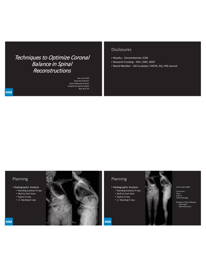
Planning

  • Radiographic Analysis
  • Standing Scoliosis X-rays
  • Skull to Foot View
  • Supine X-rays
  • +/- Bending X-rays

Planning

  • Radiographic Analysis
  • Standing Scoliosis X-rays
  • Skull to Foot View
  • Supine X-rays
  • +/- Bending X-rays

Assess Leg Lengths Concurrent Hip or Knee or Ankle Pathology Etiology of Pelvic Obliquity

  • Leg Lengths
  • Deformity Driven
slide-2
SLIDE 2

Planning

  • Radiographic Analysis
  • Standing Scoliosis X-rays
  • Skull to Foot View
  • Supine X-rays
  • +/- Bending X-rays

Flexibility Assessment

Osteotomy Selection Osteotomy Selection

  • SPO 11°
  • PSO 33°
  • Grade 4 Schwab 40°
  • Lumbar
  • Sagittal Deformities
  • VCR 50-70% Correction
  • Thoracic
  • Acute/Angular Deformities
  • >100 Deg

Relationship of PI to Lordosis

8

45° 83° 45° 20° 45° 50° 80 80° 30°

Low PI S1 ~ horizontal Flat Lordosis Large PI S1 is tilted

slide-3
SLIDE 3

Relationship of PI to Lordosis

9

45° 83° 45° 20° 45° 50° 30 80° 30°

Low PI S1 ~ horizontal Flat Lordosis Large PI S1 is tilted

Distribution of Lumbar Lordosis versus PI

  • On average, distal lordosis ~60%
  • f the total lordosis
  • Value almost constant across PI

group

  • Significant association between

proximal lordosis and PI

  • Larger value
  • More lordotic levels

10 p<0.001 p<0.001 p=0.03

Goals of the Surgery

  • Coronal and Sagittal Balance
  • Not Necessarily Cobb Angle!
  • +/- Decompression
  • Fusion
  • Minimize EBL and Intra-op

Complications

  • Avoidance of Neurologic

Complications

  • Starts with Planning!

Case Example

  • 60F with Back Pain
  • Coronal and Sagittal

Decompensation

  • PI: 45
  • LL: +50 Kyphosis
  • PT: 35 deg
  • Coronal Cobb: 66
  • SVA +15 cm
slide-4
SLIDE 4

Supine X-rays

  • Element of Rigidity
  • Will need Osteotomy
  • CT – no Fusions
  • MRI – no Stenosis

MRI

  • Element of Rigidity
  • Will need Osteotomy
  • CT – no Fusions
  • MRI – no Stenosis

Thinking Process

  • Lowest Instrumented Vertebrae
  • S1
  • S1/Ilium
  • L5
  • Upper Instrumented Vertebrae
  • UT (T2, T3, T4)
  • LT (T9, T10)

Thinking Process

  • Lowest Instrumented Vertebrae
  • S1
  • S1/Ilium
  • L5
  • Upper Instrumented Vertebrae
  • UT (T2, T3, T4)
  • LT (T9, T10)
  • Osteotomy Selection
  • SPO?
  • PSO?
  • VCR?

T3

slide-5
SLIDE 5

Getting the Balance

  • SPOs T11-L4
  • Intra-op 36” Cassette
  • Still with Significant Left

Coronal Decompensation

Iliac Screw to “Titrate” Coronal Balance

Distraction until the Pelvis is Leveled Then Place Right Sided Rod and Final Tighten – Locks in Orientation

LL: 32 deg SVA: +5 cm PT: 15

Coronal Plane can be Tricky

  • 40F Left Leg

Pain and Back

  • Right LLD

by 3cm

  • Correction

must plan for this

98

slide-6
SLIDE 6

98 94

PATIENT HISTORY

  • A

Age: 52 yrs

  • Gender: Female
  • Functional Status: Fully ambulatory

CC: Leftward tilting posture and back pain HPI: 52F who presented after thoracolumbar fusion in 2010

for AIS diagnosed as a teen followed by 2013 revision for bilateral rod After the revision, gradually increasing leftward postural tilt with significant back painbreakage at the distal aspect of her construct. She had no bowel/bladder issues, no radicular pain and no numbness/weakness.

PSHx:

  • 2010 T3-Pelvis fusion
  • 2013 Spinal fusion revision

with removal of hardware

PRE-OPERATIVE IMAGING- 2010

Coronal T1 - T5: 26 T5 -T10: 59 T10 – L4: 57 Sagittal T2-T12: 39 L1-S1: 56 Coronal Benders T1 - T5: 26 -> 9 T5 -T10: 59 -> 49 T10 – L4: 57 -> 17

slide-7
SLIDE 7

PRE-OPERATIVE IMAGING PRE

RE RE RE RE RE RE RE RE RE RE RE RE RE RE RE RE RE RE RE RE RE RE RE RE RE RE RE RE RE RE RE RE E RE RE RE E RE RE RE RE RE E RE RE RE RE RE RE RE E RE RE RE RE RE RE RE R -O

P

E IMA MA A MA A MA MA A MA MA A MA MA A MA MA A MA MA A MAGI GI GI GI GI GI GI GI GI GI GI GI GI GI GI GI GI GI I GI GI GI I GI GING NG NG NG NG NG NG NG NG NG NG NG NG NG NG NG NG PE PE PE PE PE PE PE PE PE PE E PE E PE PE PE PE PE PE PE PE PE E PE ERA RA RA RA RA RA RA RA RA RA RA RA RA A RA RA RA RA RA RA RA RA RA RA A RA RA RA RA A RA RA RA RA A RA RA RA RA RA RA A RA RA RA RA RA RA RA RA RA RA A RA RA R T P TI TI TI TI TI TI TI TI TI TI TI TI TI TI TI TI TI TI TI TI TI TI I TI TI TI TI TI TI TI TIVE V

71mm SVA 59d PI 31d PT 28d SS 30d LL PI-LL = 29

PRE-OPERATIVE IMAGING PRE-OPERATIVE IMAGING POST-OPERATIVE IMAGING

slide-8
SLIDE 8

2yr f/u 2yr f/u /u /u /u u

68F

  • Back pain
  • Feels like she is leaning forward
  • No leg pain
  • H/O PSF T4-Pelvis (2010)
  • High Grade Spondy
slide-9
SLIDE 9

What would you do?

  • Surgical Options
  • 3-Column Osteotomy?

Where?

  • Distal Fixation/Levels?
  • Proximal Levels?
slide-10
SLIDE 10

T2-Pelvis Revision with S1 PSO

THANK YOU

  • hanjokimmd@gmail.com