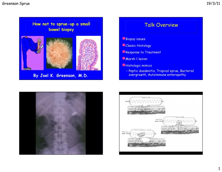
talk overview
play

Talk Overview malabsorption bowel biopsy Biopsy issues Classic - PowerPoint PPT Presentation

Greenson Sprue 19/3/11 The surgical pathology of How not to sprue-up a small Talk Overview malabsorption bowel biopsy Biopsy issues Classic Histology Response to Treatment Marsh 1 lesion Histologic mimics Peptic


  1. Greenson Sprue 19/3/11 The surgical pathology of How not to sprue-up a small Talk Overview malabsorption bowel biopsy � Biopsy issues � Classic Histology � Response to Treatment � Marsh 1 lesion � Histologic mimics – Peptic duodenitis, Tropical sprue, Bacterial overgrowth, Autoimmune enteropathy By Joel K. Greenson, M.D. * 1

  2. Greenson Sprue 19/3/11 Where to Biopsy? How many Biopsies? � Most studies suggest that 4 biopsies are optimum – One study suggested 5 with one biopsy being from the bulb � Recent pediatric studies have found 10% of kids have involvement in the bulb only and that 10% have non- diagnostic findings in the bulb with Marsh 3 lesions more distally. � Probably best to biopsy both bulb and distal duodenum and put in separate jars Weir DC Am J Gastroenterol 105;207-12:2010. Rashid M. BMC Gastroenterol 9;78:2009. 2

  3. Greenson Sprue 19/3/11 3

  4. Greenson Sprue 19/3/11 Cause of Celiac Disease Disorders of Malabsorption Wheat Flour Classification Starch � Normal mucosal histology Fat Fiber � Non-specific inflammatory and Protein architectural changes Water Insoluble Water Soluble � Demonstrable infectious agents Fraction Fraction � Immunodeficiency present Gluten � Misc. entities with characteristic findings Alcohol Insoluble Alcohol Soluble Glutenin Gliadin 4

  5. Greenson Sprue 19/3/11 Celiac Disease Histopathology - prior to Tx � Flat biopsy with surface damage � Increased Intraepithelial lymphocytes � Increased lamina propria inflammation – Plasma cells � Increased crypt mitoses 5

  6. Greenson Sprue 19/3/11 Classification of Celiac Lesions Marsh 3A Marsh 3B Marsh 3C 6

  7. Greenson Sprue 19/3/11 Gluten Free Diet - 2 Weeks Celiac Disease Histopathology - Shortly after Tx � Marked clinical improvement � Surface epithelium restored � Slight return of villi � Other findings unchanged Celiac Disease Histopathology - Long term Tx � Continued clinical improvement � Further return of villi � Mitotic rate subsides � Chronic inflammation subsides 7

  8. Greenson Sprue 19/3/11 Celiac Disease Gluten Challenge � Epithelial lymphocytes increase � Epithelial damage to upper villi � Full-blown lesion develops later Celiac Disease Pathogenic Factors � Genetic Aspects – Familial Occurrence (11-22% first degree relative) – Identical Twin Concordance (70%) – HLA Associations ( DQ2, B8) � Environmental Factors – Dietary Gluten – Twin non-concordance rate of 30%; separate onsets – ?Viral exposure (Adenovirus type 12) 8

  9. Greenson Sprue 19/3/11 Serologic Markers Protein Sequence Homology In Celiac Disease Marker Sensitivity Specificity Anti-gliadin 31-100% 85-100% Anti-reticulin 42-100% 95-100% Anti-endomysium 60-100% 95-100% Tissue Transglut 85-100% 92-97% Schuppan et al. Gastro 2009;137:1912-33 Schuppan D. Gastroenterol 2000:119;234-242 Pinier et al, Am J Gastroenterol 105:2551-2561;2010 9

  10. Greenson Sprue 19/3/11 CD Iceberg ___________________________ CD Diseased ___________________________ mucosa DQ2 latent ___________________________ Normal mucosa normal 10

  11. Greenson Sprue 19/3/11 How many IELs are abnormal? � >25/100 epithelial cells � >40/100 epithelial cells � >12/20 epithelial cells on the tips of villi – Decrescendo pattern is normal – Diffuse pattern is abnormal – Goldstein Am J Clin Pathol 116;63-71,2001 � >8/20 epithelial cells in the tips of villi – CD3 stains – Biagi et al J Clin Pathol 57;835-839, 2004 Normal Architecture But what does it all mean? Increased IELs � 2-3 % of small bowel biopsies have normal architecture � Gluten Sensitive Enteropathy – Early type 1 lesion or treated sprue with increased IELs � Other food hypersensitivity � Autoimmune conditions (RA, SLE, MS, Graves, Hashimoto ’ s, � Depending on the type of study and the country the � H. Pylori (usually only in bulb) study was carried out in, anywhere from 9 to 40% of Diabetes) such cases represent (pre) celiac disease. � Post-infection – Whether such patients need any therapy is � Drugs (NSAIDs, PPIs??) � Bacterial Overgrowth controversial � Crohn ’ s disease and Ulcerative colitis � Obesity Brown I,et al. Arch Pathol Lab Med 130;1020-25, 2006 11

  12. Greenson Sprue 19/3/11 Diseases Associated with Marsh 1 Lesions Results Celiac Disease Complications Other, 7 IBS, 9 CD, 19 � Refractory Celiac Disease H. pylori, 7 � Ulcers of Small Bowel Bacterial � Collagenous Sprue Overgrowth, 7 Idiopathic, 31 � Malignancy Crohn's, 7 – T cell Lymphoma of gut and regional nodes NSAID, 17 – Adenocarcinoma of small bowel – Squamous cell carcinoma of esophagus and Other Diagnoses: Graft versus Host Disease, Combined Variable oropharynx Immunodeficiency, Diabetes mellitus 1, Juvenile Rheumatoid Arthritis, Systemic Lupus Erythematosis, Tropical Sprue, Ulcerative Colitis Refractory Celiac Disease Refractory Celiac Disease � Develops in about 5% of celiac patients � IELs in Celiac disease and type I RCD are CD3 + and CD8 + – Malabsorption, diarrhea, pain, wt loss � Divided into types I and II � IELs in type II RCD are CD3 + and CD8 - � Type I RCD: IELs are normal / not clonal – better prognosis – Will have T-cell gene rearrangements – Can progress to Type II – Will also loose staining for T-cell � Type II RCD: IELs are aberrant / clonal receptor αβ – 50% mortality rate 12

  13. Greenson Sprue 19/3/11 Small Intestinal Ulcers In Celiac Disease CD3 CD8 13

  14. Greenson Sprue 19/3/11 LYMPHOMA IN CELIAC DISEASE 14

  15. Greenson Sprue 19/3/11 Malabsorption Sprue-like Changes Gluten Free Diet Response No Response (Refractory Sprue) Remain Well Deterioration Benign Ulcer Refractory Celiac Disease Lymphoma Celiac Disease Histologic Mimics � Celiac-related – Lymphoma (EATCL) – Collagenous Sprue � Other luminal antigens other than gluten/gliadin – Soy protein � General – Peptic duodenitis – Tropical Sprue, Bacterial overgrowth – Crohn ’ s disease – Autoimmune enteropathy – Infections/immunodeficiencies 15

  16. Greenson Sprue 19/3/11 Tropical Sprue � Chronic malabsorption after infectious diarrhea commonest in tropical regions � Bacterial overgrowth with B-12 and Folate deficiencies - often responds to antibiotics and vitamin supplements � Biopsy findings are variable – Sprue-like changes with less intense damage than full blown celiac disease – Both Jejunum and Ileum involved 16

  17. Greenson Sprue 19/3/11 Stasis Syndrome � Crohn ’ s Disease (Bacterial Overgrowth) � Diverticular Disease � Scleroderma � Pseudo-obstruction � Post-Surgical – Blind loop or Pouch – Entero-enterostomy – Afferent loop – Fistulae - Adhesions/partial obstruction 17

  18. Greenson Sprue 19/3/11 Bacterial Overgrowth Biopsy Findings � Irregular Villi � Surface cell damage � Plasmacytosis � Neutrophils � “ Doesn ’ t fit ” � Crypt Hyperplasia 18

  19. Greenson Sprue 19/3/11 Autoimmune Enteropathy � Childhood onset - usually prior to age 1 – Intractable diarrhea not relieved by TPN – Anti-enterocyte antibodies (requires indirect immunofluorescence) – Other autoantibodies (islet cell, parietal cell) – FOXP3 mutation – X-linked with polyendocrinopathy � Also Adult onset (not well known) – Anti-enterocyte antibodies or anti-goblet cell antibodies 19

  20. Greenson Sprue 19/3/11 It ’ s the Surgeon ’ s Fault! Pouchitis and Diversion Colitis 20

  21. Greenson Sprue 19/3/11 The Pouch The Pouch Baseline Histology � Increase in lamina propria chronic inflammation – Lymphs, plasma cells, eos and histiocytes � Variable villous atrophy with crypt hyperplasia mucins over time - “ Colonisation ” � Switch from small intestinal to colonic type Pouchitis 21

  22. Greenson Sprue 19/3/11 Pouchitis Pouchitis Clinical Symptoms Clinical Syndromes � Occurs in 7-50% of pouch patients (avg. 32%) � Responsive to Antibiotics (Flagyl) � Abdominal pain and fever – Bacterial overgrowth � Refractory Pouchitis � Bloody stools, increased frequency, incontinence – Irritable pouch syndrome - no path changes � Often responds to antibiotics – Short strip pouchitis - UC in retained rectal � 10-20% refractory to therapy -?chronic IBD mucosa – Much more common in UC than FAP patients – Chronic primary refractory pouchitis - active – Highest incidence in UC patients with PSC inflammation in bxs. Pouchitis Histopathology of the “ baseline pouch ” and these changes do not � Villous blunting and chronic inflammtion are part correlate with clinical symptoms � Active inflammation does correlate with clinical symptoms – Erosions – Ulcers – Cryptitis 22

  23. Greenson Sprue 19/3/11 The End 23

Download Presentation
Download Policy: The content available on the website is offered to you 'AS IS' for your personal information and use only. It cannot be commercialized, licensed, or distributed on other websites without prior consent from the author. To download a presentation, simply click this link. If you encounter any difficulties during the download process, it's possible that the publisher has removed the file from their server.

Recommend


More recommend