
SLIDE 1
Structure and Interactions of Translesion Synthesis DNA Polymerases
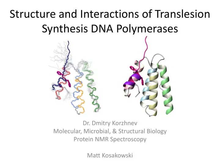
- Dr. Dmitry Korzhnev
Molecular, Microbial, & Structural Biology Protein NMR Spectroscopy Matt Kosakowski
Structure and Interactions of Translesion Synthesis DNA Polymerases - - PowerPoint PPT Presentation
Structure and Interactions of Translesion Synthesis DNA Polymerases Dr. Dmitry Korzhnev Molecular, Microbial, & Structural Biology Protein NMR Spectroscopy Matt Kosakowski What are TLS DNA Polymerases? They are a key component of DNA
Molecular, Microbial, & Structural Biology Protein NMR Spectroscopy Matt Kosakowski
Bulk DNA Replication Replicative B-family: Pol δ, Pol ε Translesional Synthesis TLS Y-family: Rev1, pol η, pol ι, pol κ TLS B-family: Pol ζ
every 106 to 108 base pairs.
DNA lesions.
ineffective in highly damaged DNA
10 to 1000 base pairs.
lesions
DNA, however is mutagenic and can incorporate new mutations in the DNA through mismatch base pairs.
interaction with PCNA, a ring shaped protein involved in replication, that otherwise serves as an activator and binding platform.
164 at a stalled replication fork, replicative polymerases are exchanged for TLS polymerases.
DNA damage tolerance pathways and PCNA ubiquitination
Pol ζ/rev1 TLS branch accounts for 90% of mutations introduced in genomic DNA.
Rad18 (Rad6) SHPRH (Mms2/Ubc13) HLTF (Mms2/Ubc13) Interactions between proteins involved in TLS
Determining a detailed, structural characterization aimed at understanding these mechanisms is our goal.
interaction with each other and with PCNA. We can find the specific amino acid sequence of these domains using protein NMR spectroscopy.
Domain architecture of TLS polymerases
– “Fingerprint” of a protein – Each peak represents an amino acid – Can involve different techniques and methods depending on size of the protein sample and resonance assignments. – Use of N15 and C13 labeled protein to map chemical shifts, labeled amine groups resonate at a different frequency – Requires highly purified protein grown on isotopic media.
6.0 7.0 8.0 9.0 10.0 108 114 118 122 126 130 134 110
1H [PPM] 15N [PPM]
NH TROSY - scPCNA (100 kDa)
hPAD FPLC Gel Fractions hPAD HSQC Spectrum
1H 15N
1 2 3 4 5 6 7 8 9 10 ~20 KDa