
SLIDE 1
Structure and assembly of bacterial pili, from analysis of virulence-related gene clusters
Ted Baker
University of Auckland New Zealand
Structure and assembly of bacterial pili, from analysis of - - PowerPoint PPT Presentation
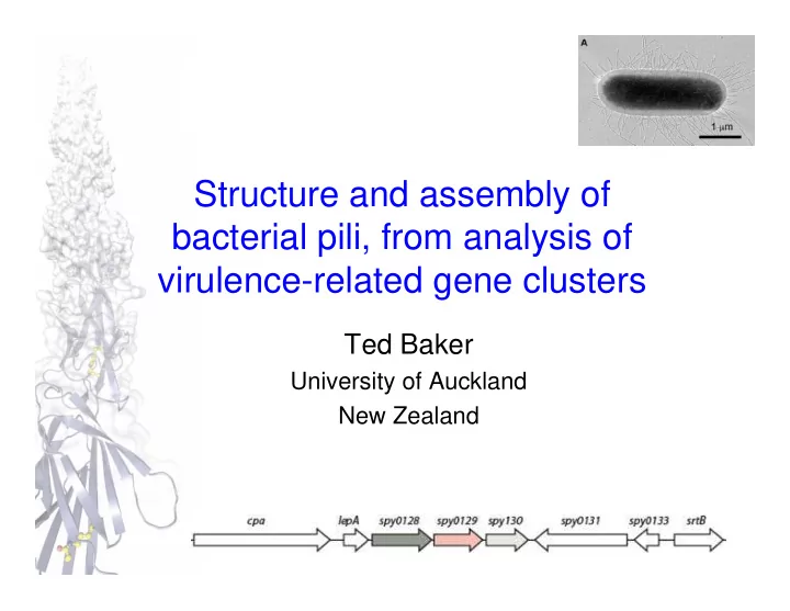
Structure and assembly of bacterial pili, from analysis of virulence-related gene clusters Ted Baker University of Auckland New Zealand Context: virulence-associated proteins from Streptococcus pyogenes Serious human pathogen mild skin
University of Auckland New Zealand
mild skin and throat infections serious invasive disease
wide sequence variation
Spy0125 (cpa, collagen binding) VPPTG Spy0128 (unknown function) EVPTG Spy0130 (unknown function) LPxTG
M1
Sortase substrates
Other genes
genes)
assembled into filaments
pili from Gram-negative bacteria
Neisseria
available and predicted surface proteins could be expressed.
Å)
gold-labelled antibodies
gene deletions:
and extremely stable and protease-resistant
linking C-terminus to NH2 group of peptidoglycan
to NH2 group of a Lys residue on next covalent polymer
Spy0129 HaeJoo Kang
two Ig-like domains
~10 nm ~2-3 nm
N-domain C-domain
packing
(like actual pili)
in position to join to C-terminus of next subunit and generate extended pilus – Lys161 (if our crystal packing model was correct!)
HaeJoo Kang Fiona Clow Martin Middleditch
There are known knowns. These are the things that we know that we know. There are known unknowns. That is to say, there are things that we know we don’t know. But there are also unknown unknowns. These are things we don’t know we don’t know.
Rumsfeld, D. (2002). Press briefing.
joining Lys and Asn side chains
Isopeptide bonds Spontaneously formed
One in each domain Catalytically-essential Glu residue FctB Cpa Kang et al. (2007) Science 318 1625-1628
Lys NH2 Asn H2N O
Lys H N Asn O NH3
Basal subunit Adhesin at the tip
for incorporation into pilus
collagen-like helix reaches into cell membrane?
FctB Cpa BP BP Christian Linke Paul Young JBC (2010) 285, 20381-20389
link FctB to the shaft
and is essential for adhesion
between Cys and Gln side chains
Covalent binding to host cells?
FctB BP BP
Pointon et al (2010) JBC 285 33858- 33866
not easily identified by sequence analysis)
2 domains
3 domains
4 domains
unrecognized isopeptide bonds in cell matrix-binding surface proteins of Gram-positive bacteria
Cna from S. aureus – Collagen binding Cna A domain – Structural Cna B domain
Cna
A B B B Cna A Cna B Minor pilin from S. agalactiae
Either
(S. pyogenes, S. pneumoniae, S. agalactiae,
Or
multi-domain proteins
(from S. aureus, S. suis, S. gordonii,
stability and mechanical strength
Kang & Baker (2011) Trends in
unexpected features of protein structure
attachment)