stroke does occur in children
play

Stroke does occur in Children: Children Incidence: 1 per 3,500 - PowerPoint PPT Presentation

2/10/2017 Patient X: History Part 1 Recent Advances in Previously healthy 14-year old boy Neurology While playing basketball with friends, he had a witnessed convulsion lasting six minutes. Difficult Cases Afterwards he had a mild


  1. 2/10/2017 Patient X: History Part 1 Recent Advances in • Previously healthy 14-year old boy Neurology • While playing basketball with friends, he had a witnessed convulsion lasting six minutes. Difficult Cases • Afterwards he had a mild headache and right- Heather J. Fullerton, MD, MAS sided weakness. Professor of Neurology & Pediatrics • He was transported by EMS to a hospital where Director, Pediatric Brain Center he was found to be aphasic with a right hemiparesis. 2 DDX for acute hemiparesis in a child 
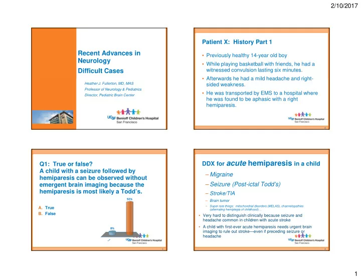
Q1: True or false? A child with a seizure followed by – Migraine hemiparesis can be observed without – Seizure (Post-ictal Todd’s) emergent brain imaging because the hemiparesis is most likely a Todd’s. – Stroke/TIA 92% – Brain tumor – Super rare things: mitochondrial disorders (MELAS), channelopathies A. True (alternating hemiplegia of childhood)… B. False • Very hard to distinguish clinically because seizure and headache common in children with acute stroke • A child with first-ever acute hemiparesis needs urgent brain 8% imaging to rule out stroke—even if preceding seizure or headache e e s u l r a T F 3 4 1

  2. 2/10/2017 Half of strokes in kids are hemorrhagic Stroke does occur in Children: Children • Incidence: – 1 per 3,500 neonates – 4.6 per 100,000 children/year in US Adults Ischemic Hemorrhagic Hemorrhagic – About 5,000 US kids/year Ischemic Broderick, J Child Neuro, 1993 Agrawal, Stroke, 2009 Approach to Imaging for Suspected Stroke Approach to Imaging • What about CT/CTA? • Emergent MRI with DWI/ADC – Use sparingly: radiation more concerning in young • Followed by kids immediate brain MRA if + infarct – Useful if MR can’t be done • Especially if (pacer leads) or can’t be within 6 hour done quickly & thrombectomy thrombectomy would be window considered 10 hr old infarct in 5 month old • More thorough – Sensitive for hemorrhage, with congenital heart dz vascular imaging not acute infarct later – Don’t add CT perfusion (not worth the radiation) 2

  3. 2/10/2017 Q2: What is the most common cause of Patient X: History Part 2 arterial ischemic stroke in a previously healthy child? Back to our 14 year old boy with a convulsion followed by right hemiparesis and aphasia… A. Embolism from congenital heart disease • Emergent MRI: small infarcts 41% B. Embolism from endocarditis of L MCA territory 34% C. Sickle cell disease • MRA: mild narrowing of his left supraclinoid internal D. Genetic thrombophilia 17% carotid artery (ICA)— E. Arteriopathy thrombus versus 5% 2% arteriopathy? e s a y . i s i h . . t a l a i h i t d e a t r s p p i a i n d o o e c o b r i g l l m e n d e n c o t o r c e e r A h m m l k t c o o i c r r S i f f t e m m n e s s G i i o l o l b b m m E E 9 10 Kids with arteriopathy are at highest risk of recurrence Arteriopathy (disease of a cervical or 1.00 Proportion recurrent stroke free cerebral vessel) is the most common cause Almost 0.75 of childhood arterial ischemic stroke…. 1 in 4 0.50 And the strongest predictor of recurrent 0.25 stroke . 0.00 0 3 6 9 12 15 18 21 24 Fullerton, Time from index AIS to first recurrent stroke, months Stroke , Idiopathic 2015 Cardioembolic Possible arteriopathy Definite arteriopathy 11 3

  4. 2/10/2017 Patient X: History Part 3 MRA on transfer • Admitted to the hospital and placed on IV heparin. An echocardiogram was normal. His deficits improved dramatically over the next few days. • 3-days post stroke: severe left frontal headache. • 4-days post-stroke: agitated with worsened aphasia and right hemiparesis. • His deficits were positional, improving when placed flat, and responded to IV hydration and pressors. • Repeat MRI/A: new infarction in the left MCA territory and severe narrowing of his left distal ICA and proximal MCA . • Transferred to UCSF 13 14 FCAi Q3: What causes rapidly progressive stenosis of the distal ICA in children? Beading of M1 on conventional angiography Lenticulostriate infarcts A. Focal Cerebral Arteriopathy—Inflammatory (FCA-i), also known as Transient Cerebral Arteriopathy (TCA) B. Moyamoya disease (idiopathic moyamoya) C. Moyamoya syndrome (secondary moyamoya) 62% D. Kawasaki disease 18% 14% 5% e s . . . . a . . . . . e p o s o i ( i s d d r i i e e ( m i e k r t s o a A a s r a l e d a s n w r i a b d y s K e a a r y e o y C o m m l a a a c y o o y F M o M 15 16 4

  5. 2/10/2017 Natural History of FCA-i What causes FCA-i? • Monophasic disease • Varicella zoster virus (VZV)— strong evidence from histopathology studies • Initial progression • Other herpesviruses, like herpes simplex virus, type 1 • Nonprogression after (HSV-1)— evidence from case reports and a prospective case- 6 months control study (Vascular effects of Infection in Pediatric Stroke, VIPS study) • Improvement or stabilization; rarely • Other pathogens? normalization • Abnormal host immune response? – Chabrier & Sebire, J Child Neurol 1998; Danchaivijitr, Acute 2 months 12 months Ann Neurol 2006 Courtesy of G. DeVeber 18 Q4: What are proven therapies for FCAi? Patient X: History Part 4 • Admitted to the PICU, placed on IVF (1.5 x maintenance), head A. Aspirin of bed flat, on IV pressors B. Revascularization Surgery (STA-MCA bypass) • Had flow-dependent deficits: when upright, hemiparesis C. Acyclovir worsened 37% D. Corticosteroids • Serologies positive for HSV-1 E. None of the above • After several days of observation, with no improvement in flow- 23% dependent deficits, treated with IV corticosteroids and acylovir 18% 13% • After several days, patient stabilized 9% • Slowly weaned off IVF and pressors • 2 month follow-up: dramatic improvement in ICA stenosis n r s e i . i d r . v v i . o i o p y o r l r b s e c e a A y g c t r s e • 2 year follow-up: no further strokes u A o h S c t i n t f r o o o i e t C a n o z i N r a l u c s a v e R 19 20 5

  6. 2/10/2017 Before his arteriopathy progressed, Follow-up MRA could we have diagnosed him with FCAi? • FCA differential diagnosis: – FCAi – FCAd (focal cerebral arteriopathy, dissection subtype) • Intracranial dissection • Can also show early progression (spiral dissection) • Difficult to distinguish from FCAi • Vessel Wall Imaging (VWI) can help – Early, unilateral moyamoya • Lenticulostriate collaterals • Chronic, silent infarcts 21 22 Moyamoya disease/syndrome Enhancement on VWI: suggests FCAi • Slow narrowing of bilateral supraclinoid ICAs • 2ary lenticulostriate collaterals • Can be unilateral at onset • Secondary form: sickle cell disease, Down syndrome, NF-1, cranial radiation for cancer Courtesy of Max Wintermark, MD 6

  7. 2/10/2017 Childhood arteriopathy diagnosis important for management…and prevention of recurrent stroke Aspirin Heparin Revascularization Activity Hydration & Surgery (EDAS, Restriction BP STA-MCA bypass) augmentation FCAi x Rarely Acutely Dissection x Sometimes Lifelong Moyamoya x Routinely Long term Clinical trials planned for corticosteroids and acyclovir for FCAi. P.S. FCAi has also been reported in young adults 25 7

Download Presentation
Download Policy: The content available on the website is offered to you 'AS IS' for your personal information and use only. It cannot be commercialized, licensed, or distributed on other websites without prior consent from the author. To download a presentation, simply click this link. If you encounter any difficulties during the download process, it's possible that the publisher has removed the file from their server.

Recommend


More recommend