
SLIDE 1
Intervet symposium om Kvarka. Lund 2005-03-16. Stockholm 2005-03-17. Föreläsare: Josh Slater, Royal Veterinary College, University of London
1
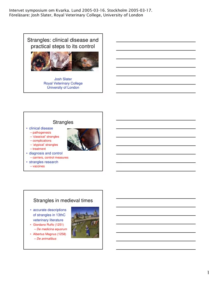
Strangles: clinical disease and practical steps to its control
Josh Slater Royal Veterinary College University of London
Strangles
- clinical disease
– pathogenesis – ‘classical’ strangles – complications – ‘atypical’ strangles – treatment
- diagnosis and control
– carriers, control measures
- strangles research
– vaccines
Strangles in medieval times
- accurate descriptions
- f strangles in 13thC
veterinary literature
- Giordano Ruffo (1251)
– De medicina equorum
- Albertus Magnus (1258)