
SLIDE 1
11/8/2013 1
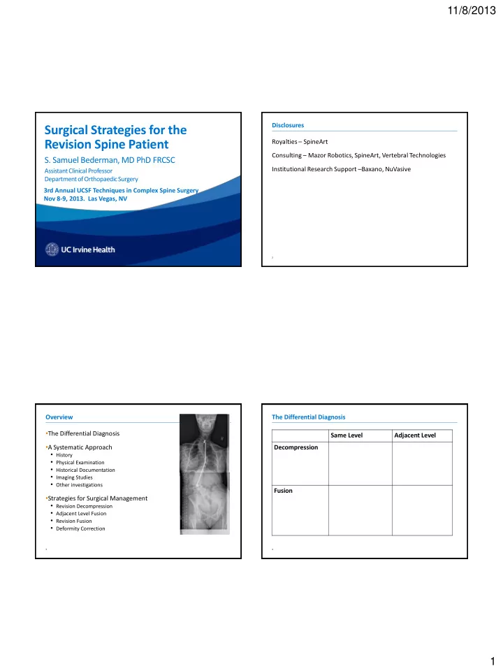
Surgical Strategies for the Revision Spine Patient
- S. Samuel Bederman, MD PhD FRCSC
Assistant Clinical Professor Department of Orthopaedic Surgery 3rd Annual UCSF Techniques in Complex Spine Surgery Nov 8-9, 2013. Las Vegas, NV
2
Disclosures Royalties – SpineArt Consulting – Mazor Robotics, SpineArt, Vertebral Technologies Institutional Research Support –Baxano, NuVasive
3
Overview
- The Differential Diagnosis
- A Systematic Approach
- History
- Physical Examination
- Historical Documentation
- Imaging Studies
- Other investigations
- Strategies for Surgical Management
- Revision Decompression
- Adjacent Level Fusion
- Revision Fusion
- Deformity Correction
4
The Differential Diagnosis Same Level Adjacent Level Decompression
Infection CSF leak Stenosis Fracture Instability/Deformity Stenosis
Fusion
Infection Stenosis Fracture Symptomatic Hardware Pseudarthrosis Instability/Deformity Stenosis Fracture Instability/Deformity