Random Conical tilt 3D reconstruction Central section theorem Tilt - - PDF document

random conical tilt 3d reconstruction
SMART_READER_LITE
LIVE PREVIEW

Random Conical tilt 3D reconstruction Central section theorem Tilt - - PDF document

Sali A, Glaeser R, Earnest T, Baumeister W. (2003) From Words to literature in structural proteomics. Nature 422 (6928): 216-225. Random Conical tilt 3D


slide-1
SLIDE 1

Random Conical tilt 3D reconstruction

  • Central section theorem
  • Euler angles
  • Principle of conical tilt series
  • Missing cone artefact
  • Multivariate statistical analysis
  • Early 3D studies and negative staining problems
  • Perspectives and new trends

Nicolas Boisset CNRS Département de Biologie Structurale Institut de Minéralogie et de Physique des Milieux Condensés I.M.P.M.C. UMR 7590 CNRS, Université Pierre & Marie Curie 140 Rue de Lourmel, 75015 Paris email: nicolas.boisset@impmc.jussieu.fr

Tilt series Back-projection & 3D reconstruction

Sali A, Glaeser R, Earnest T, Baumeister W. (2003) From Words to literature in structural proteomics. Nature 422(6928): 216-225.

2 5 4 3 6 7 1

In reciprocal space, every 2D projection of a 3D object corresponds to a central section in the 3D Fourier transform of the

  • bject. Each central section is orthogonal

to the direction of of projection.

Central projection theorem

1 2 7 6 5 4 3

Constraints of cryoEM on biological objects : Work with low electrondose (~10e-/Å2) => the less exposures, the better. Images have a low signal-to-noise ratio Compromise defocus level with resolution (CTF) Computing 2D or 3D numeric averages (only one conformation assumed in the sample) Use internal symmetries of the objects : helicoïdal symmetry, icosaedral symmetry, 2D crystals, or no symmetry at all…

X y Z

  • X

Y Z

Euler angles

Convention for Euler angles in SPIDER Phi Theta Psi

1 2 5 4 3 6 7 8

0° 45°

1 2 8 7 6 5 4 3

With only two exposures a conical tilt series can be generated

Angular distribution represented on a spherical angular map Radermacher, M., Wagenknecht, T., Verschoor, A. & Frank, J. Three- dimensional reconstruction from a single-exposure, random conical tilt series applied to the 50S ribosomal subunit of Escherichia coli. J Microsc 146, 113-36 (1987).

slide-2
SLIDE 2

1 2 5 4 3 6 7 8

Reciprocal space Principle of random conical tilt series

Z

Radermacher, M. (1988) Three-dimensional reconstruction of single particles from random and nonrandom tilt series. J Electr.Microsc. Tech. 9(4): 359-394. You just need to determine de Euler angles specific to each tilted-specimen image. = 0° = 90°

Interactive particle

  • selection. Picking of

image paires (45° & 0°) provides a mean to compute the :

  • direction of tilt axis ()
  • and the tilt angle ().

= Tilt angle => COS ( ) = d / D (but you don’t know if it is + or – d d D D X Y Z = in-plane direction of tilt axis If parallel to axis Y, then = 0°

0° 45° Interactive windowing at 0° and 45° tilt

2D projections are identical, except for an in plane rotation corresponding to Euler angle . 2D projections are not identical due to the tilt. Moreover, neighboring particles start to overlap

2D alignment of untilted-specimen images and computation of angle Centering and masking of tilted- specimen images

Rotation of each 0° projection by its -angle A circular mask hides (up to a certain point) the neighboring particles.

Simple back-projection Reciprocal space half-volume

=36° =36°

Once the 3 Euler angles are determined, the 3D reconstruction can be performed from the tilted- specimen projections. The simple back-projection is nothing more than adding in reciprocal space the FT of the 2D projections in their relative orientations (waffle-like distribution of central sections), followed by Fourier transform of this 3D distribution to come back in real space.

Uneven distribution of the signal Why does it look so bad?

Weigthed back-projection Reciprocal space half-volume

=36° =36° Missing cône Similar as previously, but after applying a band-pass filtering or R* weighting of the signal (lowering contribution in low spatial frequencies). It is better, but we have a non- isotropic reconstruction. Why ?

slide-3
SLIDE 3

Simultaneous Iterative reconstruction techniques (SIRT) Real space & iterative:

=36° =36° In real space with iterative methods, a starting volume is computed by simple back-projection. Then, the volume is re- projected in its original directions and 2D projection maps are compared with the experimental EM images. The difference maps [(EM) minus (2D projection of the volume)] are computed and back-projected to correct the 3D reconstruction volume. To avoid “over- correcting” the structure, the 2D difference maps are multiplied by an attenuation factor (with ~ 0.5.E-04 to 0.1.E-06). This process is iterated and at each step the “global error” between EM images and the computed volume is measured to check improvement.

Correct value ?

Global error

1.0 0.01

Iterations to small correct

1 2 3

to big

The lambda value must be adjusted depending on the quality and number of images and on imposed symmetries

Original object Simple back-projection Weigthed back-projection SIRT Comparing 3D reconstruction techniques

Front views Top views

The missing cone artifact + = + =

Top views Front views

Boisset N., Penczek P., Taveau J.C., You V., de Haas F., Lamy J. (1998) Overabundant single-particle electron microscope views induce a three-dimentional reconstruction artifact. Ultramicroscopy, 74: 201-207.

even angular distribution uneven angular distributions

In rare occasions, a single overabundant preferential orientation can distort your structure when using SIRT

0° 45°

Titled-specimen 45° Untilted specimen 0°

Interactive particle picking with determination

  • f tilt axis direction () and tilt angle ()

0° 45°

slide-4
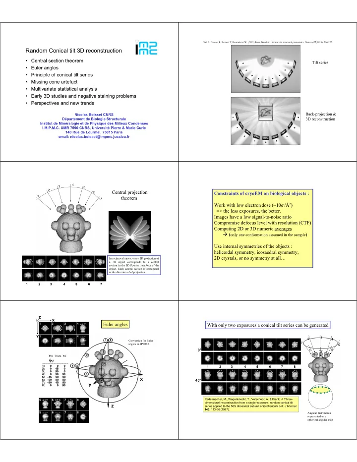
SLIDE 4

Original side views Aligned side views Alignment Determination

  • f angle (

MSA and clustering 5 views

A simplified and therefore mathematically incorrect description of Correspondence analysis (CORAN) To get the “flavour” of this method.

You have normalized, aligned a set of noisy images and you want to sort them automatically. (For correspondence analysis no negative density is tolerated, while for principal component analysis (PCA) you don’t care). 1- Create a mask following the shape of the total average 2- For each image, extract all densities from the pixels falling within the mask and re-dispose then into a line.

Image No1 Image No76 Pixel 1 Pixel 2754 Sum per colum Sum per line Total sum

K

ij

  • f. j

fI .

K

ij

3- Place theses lines of densities into a table 4- An other way to consider the data is to say that these densities are coordinates in a multidimensional space. 5- Hence in this example, each image having 2754 pixels under le mask, our data set corresponds to 76 images, that we can consider as 76 dots in a space of 2754 dimensions. “Intelligenti pauca” = intelligent people understand each other with a few words ! …

1. Absolute values frequencies Kij Kij / kij = fij

  • 2. Euclidian distance 2 distance

fij fij / fi. f.j

  • 3. Image mass i = fi.

Origine changed to the center of gravity

  • f the table = -f.j
  • 4. Diagonalization of the covariance matrix

Xij = (fij – fi. f.j) / fi. f.j equivalent to a least square fit to define new factorial axes (eigen vectors) and the coordinates of each image on these axes.

Intuitively one can guess that two identical images will have similar coordinates in the multi-dimensional space. Therefore in the multidimensional space they correspond to two dots located near each other. Conversely, two dissimilar images will correspond to two dots located far away from each other. Multi-dimensional statistical analysis (MSA), reinforces this idea of “similarity = proximity” but it changes the coordinate system of our data set in order to reduce the number of dimensions to a number a few meaningful axes. These axes or “eigen vectors” correspond to main “trends” or “variations” within our population of images.

The ALMOND approach

Space with 2754 dimensions

2 3 1

Eigen vector 1 Eigen vector 2 Eigen vector 3

One method of diagonalization of the co-variance matrix (T = X’ X), called “la méthode de la dragée” or the “Almond method” illustrates what happens at this stage. The original multi-dimensional space has been distorted by the chi square matrix to express the variations among the

  • images. Schematically one can say that the cloud of 76 dots (representing our 76 images) which was originally

contained in a multi-dimensional “sphere” is now contained within a multi-dimensional “almond”. 1. The longest dimension of the almond corresponds to the major “trend” or variation among the image set and is defined as the first eigen vector. Its amplitude (length) corresponds to the first eigen value 1. Coordinates of our 76 dots along this new axis are calculated. 2. Then, the second longest dimension of the almond, orthogonal to the first eigen vector is determined (width of the almond). This second direction corresponds to eigen vector number two and corresponds to the second variation among the images. The amplitude of this second vector is the second eigen value 2. Coordinates of

  • ur 76 dots along this new axis are calculated.

3. Then the third longest dimension of the alond, orthogonal to the first and second eigen vectors is determined (thickness of the almond). This third direction corresponds to eigen vector number three and corresponds to the third variation among the images. The amplitude of this third vector is the third eigen value 3. Coordinates of

  • ur 76 dots along this new axis are calculated.

etc…. Eigen vector 1 Eigen vector 2 The 76 dots can be projected on planes defines by two selected eigen vectors. Here again the “proximity = similarity” rule applies, and we can identify four types of images in the example set of images. In fact, the information contained in our data is so much compressed that a set of coordinates on the eigen vectors can characterize a given image. Jean-Pierre Brétaudière and Joachim Frank designed “reconstitution and importance images” to express this relationship and to explore the variation related to each eigen vectors. For example, according to you, how looks an image having for coordinates zero on all eigen vectors ? 0, 0, 0, etc..

  • 0.2, 0, 0, etc..

+0.2, 0, 0, etc..

slide-5
SLIDE 5

Brétaudière JP and Frank J (1986) Reconstitution of molecule images analyzed by correspondence analysis: A tool for structural interpretation.

  • J. Microsc. 144, 1-14.

Axis 1 Axis 2 Axis 3 Axis 4 Axis 5 Axis 6 Axis 7 Positive importance Negative importance Positive reconstitution Negative reconstitution

Classification Ascendante Hiérarchique

1 2 3 4 5 1 2 3 4 5 1 3 6 6 5 2 7 7 4 8 8

Hierarchical ascendant classification

slide-6
SLIDE 6

External surface rendering Central cavity Hémocyanine of Helix pomatia

Mur collier

a b c d e f g h Model of Mellema & Klug (1972)

Question : Which EM views would you use to suppress the missing cone while enforcing a D5 symmetry ?

If 0° tilt images = TOP views, the missing cone axis is parallel to the five- fold axis of the cylinder. If 0° tilt images = SIDE views the missing cone axis is orthogonal to the five-fold axis of the cylinder.

When enforcing symmetry D5 or C5, you will fill up the missing cone of the SIDE views, but the missing cone of the TOP views will always superpose to itself and remain empty.

3 5 6 10 11

How to merge volumes when you don’t know if your structure has symmetries ?

Poster of Magali Cottevieille on the Glutamate synthase complexe. magali.cottevieille@impmc.jussieu.fr

VCLA005 (48 images) VCLA006 (35 images) VCLA010 (48 images) VCLA011 (63 images)

Aligning the first volumes with large missing cone artifact can be challenging if you don’t enforce any symmetry. Aligned and merged Classes 5 & 6 Aligned and merged Classes 10 & 11 Aligned and merged Classes [5, 6] & [10,11] Successive merging of volumes by pairs of closely related EM views. The resulting volume was then used as a reference to align all available images (tilted and untilted-specimen images 437 x2 ). At last additional untilted-specimen images are added to the refinement process (1344 images). 437 x 2 = 874 images Plus 1344 images refinement refinement (no symmetry imposed)

slide-7
SLIDE 7

25 Å resolution volume 9.5 Å resolution volume

5 nm

More images & refinement

9.5 Å

FSC 0.5 Amplitude corrected with SAXS data

Lambert, O., Boisset, N., Taveau, J. C. & Lamy, J. N. (1994) Three-dimensional reconstruction from a frozen-hydrated specimen of the chiton Lepidochiton sp. hemocyanin. J Mol Biol 244, 640-7.

How to sort cylindrical particles with Cn symmetry ?

Lambert, O., Boisset, N., Taveau, J. C., Preaux,

  • G. & Lamy, J. N. (1995) Three-dimensional

reconstruction of the D and C-hemocyanins of Helix pomatia from frozen-hydrated specimens. J Mol Biol 248, 431-48.

The Orthogonal tilt reconstruction method Andres E. Leschziner & Eva Nogales

Two images are recorded with specimen tilts of -45° and +45°

In this case you don’t get a conical tilt but the equivalent of a 360° tomogram.

Single layer negative staining technique Use thick staining or double-layer negative staining

  • r « sandwidch » technique

Why not use negatively stained images ?

Avoid thin stain !

Small particles less than 400 kDa

  • r hardly visible

in cryoMET

a b a b

Frozen-hydrated sample Double-layer negative staining technique

slide-8
SLIDE 8

Integration of 3D Cryo-EM in MSD & other data banks

URL: http://www.ebi.ac.uk/msd/

references:

  • Random conical tilt series (RCT) :

Rader Radermacher macher, M. (1988) Three , M. (1988) Three-

  • dimensional reconstruction of single particles from

dimensional reconstruction of single particles from random and nonrandom tilt series. random and nonrandom tilt series. J J Electr.Microsc Electr.Microsc. Tech. . Tech. 9(4): 359 9(4): 359-

  • 394.

394. 1. Check the Spider web site where tutorials are available, 2.

  • r contact Michael Radermacher at University of Vermont (Burlington) .
  • SIRT and alignment of 3D volumes :

Penczek, P.A., Grassucci, R.A., Frank, J. (1994) The ribosome at improved resolution: new techniques for merging and orientation refinement in 3D cryo-electron microscopy

  • f biological

particles. Ultramicroscopy 53: 251–270.

  • Review :

Sali A, Glaeser R, Earnest T, Baumeister W. (2003) From Words to literature in structural proteomics. Nature 422(6928): 216-225.

  • Orthogonal tilt reconstruction (OTR) :

Andres E. Leschziner & Eva Nogales (2005) The Orthogonal tilt reconstruction method: an approach to generating single-class volumes with no missing cone for ab initio reconstruction of asymmetric particles.

  • J. Struct. Biol (in press).

A BIG THANK YOU to all the people involved in the

  • rganization and running of the course,

and especially to Bridget, Clint, and Ron.