
SLIDE 1
Page 1 of 14
Purchasing Permanent Slides at Low Cost Gregor Overney Page 2 of 14 - - PDF document
Page 1 of 14 Purchasing Permanent Slides at Low Cost Gregor Overney Page 2 of 14 Purchasin ing Permanent S Sli lides a at Low C Cost This short article is for anybody interested in purchasing permanent slides at lower cost. I will focus
Page 1 of 14
Page 2 of 14
Purchasin ing Permanent S Sli lides a at Low C Cost
This short article is for anybody interested in purchasing permanent slides at lower cost. I will focus primarily on the experience in the US. “I do not think it is a good investment in time and effort to make your own (probably imperfect) permanent slides. The only permanent slides that I can recommend with good conscience that students make themselves are of diatoms.” – Werner Nachtigall For most newcomers to microscopy, I tend to agree with the author of one of the best introductions to microscopy that is still available in bookstores [1]. Making permanent slides of plant and animal tissue, for example, is a “serious” hobby in itself. But I would not discourage any curious mind in attempting to embark on this task. A good booklet about slide mounting describing safe microscopic techniques suitable for most beginners was recently published by the late Walter Dioni [2]. Please keep in mind that I refer to permanent slides and not to slides commonly referred to as fresh preparations, which can, under certain conditions, last for days or even
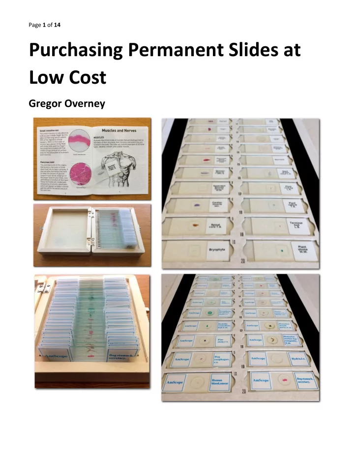
Browsing through the catalog of one of the largest online stores, I was able to find two suppliers of permanent slides at low cost (less than twenty US dollars for a set of 25 slides). From each, I purchased the 25 permanent slides set containing various biological specimens. The two suppliers are
Comparing the almost identical hard wood slide case, I expected the permanent slides to be of similar make, too. But I was in for a surprise. Photomicrographs (photographs through the microscope) are only included to give a visual impression and not to document any microscopic features. Therefore, I did not add scale bars or other annotations. The microscope
details on the production of the photomicrographs. If the illumination type is not specified, transmitted (diascopic) brightfield illumination is used. In the following, we use
through second linear polarizer (analyzer)
Page 3 of 14
AmSco cope pe – 25 Pre repare red Bi Biol
roscop
Slid ides
The following lists some of the 25 prepared slides from this set. #25. Dog stomach, secretary. 20x Plan Fluor NA 0.50. Clear tear of section. #19. Rabbit testis, sec. 20x Plan Fluor NA 0.50. This is reasonable section with good staining.
Page 4 of 14 #14. Dog esophagus, c.s. 40x Plan Fluor objective NA 0.75. This is a good histological preparation. #12. Hydrilla verticillata leaf, w.m. 20x Plan Fluor NA 0.50. This aquatic plant has sharp thorns.
Page 5 of 14 #11. Nymphaea of Aqustio (water lily) stem, c.s. 10x Plan Fluor NA 0.30. Nice preparation of an aquatic plant. #9. Lilium anther, c.s. 20x Plan Fluor NA 0.50. Good preparation.
Page 6 of 14 #7. Pumpkin stem, c.s. 20x Plan Fluor NA 0.50. Average preparation. #6. Pine leaf, c.s. 10x Plan Fluor NA 0.50. Good section.
Page 7 of 14 AmScope – 25 Prepared Biology Microscope Slides
Page 8 of 14 Quality assessment of prepared slides: Number of good quality slides: 8 Number of average quality slides: 8 Number of poor quality slides: 7 Number not graded: 2 Some impressions:
This is not suitable for a learning tool.
staining is mainly caused by old staining solution or prolonged rinsing cycle for thin sections.
most certainly not very educational. During the nineteenth century and prior to the extensive use of the famous Golgi method (chrome-silver reaction) for staining neural sections, the Reticular Theory was the most commonly held view about the organization of the nervous system. This theory assumed that the nervous system is made of a diffuse network of nerve fibers fused with each other at various points. Of course, this would be an acceptable conclusion if merely based on observing neural tissue stained with H&E. Today, we know that neurons are individual, electrically excitable cells forming the neural network. Only after the famous Spanish scientist Santiago Ramón y Cajal used and further enhanced the Golgi method, he was able to recognize this important fact. For this preparation, the Golgi method should have been used.
Page 9 of 14
Natio ional al Geogra raphi hic – Mega Bi Biol
The following lists some of the 25 prepared slides from this set. #24. Terminal bud, l.s. 10x Plan Fluor NA 0.30. Sadly it does not state of which plant. #22. Plant root tip, l.s. 10x Plan Fluor NA 0.30. Again, no mention of which plant.
Page 10 of 14 #21. Seed of Zea mays, l.s. 20x Plan Fluor NA 0.50. The optically active starch grains are shown in polarized light illumination. #19. Rhizopus nigricans, w.m. 20x Plan Fluor NA 0.50. Good preparation of black bread mold (member of Zygomycota).
Page 11 of 14 #14. Spinal cord. c.s. using chrome-silver reaction. 20x Plan Fluor NA 0.50. This is a good preparation of a spinal cord section clearly showing neurons (part of gray matter). The neurons’ axons are clearly visible with this type of
#13. Cardiac muscle, l.s. 40 Plan Fluor NA 0.75. Clearly seen are the nuclei (dark ovals) and intercalated discs (vertical zig-zag lines).
Page 12 of 14 #12. Skeletal muscle, l.s. 40x Plan Fluor NA 0.75. #8. Artery and vein, c.s. 10x Plan Fluor NA 0.30. The thick-walled oval is the artery while the thinner ones are veins.
Page 13 of 14 National Geographic – Mega Biology Set
Page 14 of 14 Quality assessment of prepared slides: Number of good quality slides: 9 Number of average quality slides: 9 Number of poor quality slides: 3 Number not graded: 4 Some impressions:
included slides and what can be learned from them. This 21 page learning guide contains numerous color photomicrographs.
Summary ry and Conc nclusi sion
Too often, but especially with the AmScope slide set, thin histology sections show numerous tears and rips. It is evident that mass production was valued higher than quality. Another observation, again mainly with the AmScope slide set, was the very faint stain, which makes it cumbersome to identify fine structures with brightfield illumination. Both sets contained at least 8 good slides that almost justify the entire purchase price when compared to the cost from one of the more reputably slide makers [4]. The National Geographic – Mega Biology Set is recommended for its better educational value and its slightly superior quality.
Refe ference ces
[1] Werner Nachtigall, Exploring with the Microscope, Sterling Publishing Co. New York, 1996. [2] Walter Dioni, Safe Microscopic Techniques for Amateurs, Onview.net Ltd., 2014. [3] Nikon Eclipse E400 with 10x, 20x, and 40x Plan Fluor objectives. Nikon Achromat/Aplanat condenser NA=1.4. Sony NEX-5N attached to a trinocular viewing head. The real image formed by the objective is projected via a 1.4x relay lens onto the CMOS image sensor. The format of the image sensor is 23.5mm by 15.6mm with 16.1
Electronic front-curtain shutter (EFCS) was enabled. Exposure was trigged via remote sensor. [4] Some reputable companies producing permanent mounts: Triarch Inc.; Ward’s Science; Carolina; Johannes Lieder GmbH, Klaus D. Kemp Microlife Services (specialized in Diatom preparations). You can find their Websites via an Internet search. [5] G. Overney. Exploration of the Human Spinal Cord. Micscape Magazine 90 (2003).