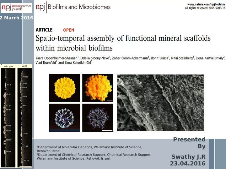
presented by
play

Presented By 1 Department of Molecular Genetics, Weizmann Institute - PowerPoint PPT Presentation

2 March 2016 Presented By 1 Department of Molecular Genetics, Weizmann Institute of Science, Rehovot, Israel 2 Department of Chemical Research Support, Chemical Research Support, Swathy J.R Weizmann Institute of Science, Rehovot, Israel.


  1. 2 March 2016 Presented By 1 Department of Molecular Genetics, Weizmann Institute of Science, Rehovot, Israel 2 Department of Chemical Research Support, Chemical Research Support, Swathy J.R Weizmann Institute of Science, Rehovot, Israel. 23.04.2016

  2. SCIENCE VOL 336 27 APRIL 2012 Fig. 1. Visualization of cyanobacterial cells forming intracellular mineral inclusions. (A) Composite epifmuorescence and optical microscopy images showing rod-shaped auto fmuorescent cyanobacteria cells. (B) Optical image shows intracellular inclusions in cyanobacterial cells (higher magnifjcation in inset). (C to F) SEM images (secondary electron mode) showing cells of the same morphotype, which systematically contain bright intracellular inclusions. (G to I) CSLM (G), phase contrast (H), and SEM (I) images of exactly the same area, showing that auto fmuorescent cells observed by CLSM contain the bright inclusions observed by SEM (inset). A difgerent, larger, and inclusion-deprived

  3. Introduction › Microbiologically induced calcium carbonate precipitation (MICP) is a bio-geochemical process that induces calcium carbonate precipitation within the soil matrix. › Calcium carbonate can be precipitated in three polymorphic forms, which in the order of their usual stabilities are calcite, aragonite and vaterite In this paper ›Granules of Calcium carbonate were observed within and on the biofjlm surfaces. ›These calcium carbonate Carbona mineral scafgolds promote te complex morphology and robustness to the biofjlms. A byproduct of metab A byproduct of metab not clearly understoo not clearly understoo Biofjlm Why this paper??

  4. Complex colony morphology correlates with calcite precipitation in Bacillus subtilis . XRD Calcite ν 2 : 875 cm -1 ν 3 : 1425 cm -1 ν 4 : 713 cm -1 (a–c) T op view of a colony of an undomesticated strain of wild-type B. subtilis (NCIB 3610). The colonies were grown on solid biomineralization-promoting medium without (a) or with (a and b) a calcium source, for 3 and 7 (a), 21 (b) days, at 30 °C, in a CO 2 -enriched environment. (b) Top view of a colony (left) and a magnifjcation of calcite crystals at the periphery (right upper) or centre (right lower) of the colony. Images were taken by Stereo microscope with an objective of × 0.5 (a and b, left) or × 1 (b, right) Scale bar corresponds to 2 mm (a and b, left) or 100 µm(b, right). (c) The FTIR spectra of calcium carbonate minerals precipitated at the edges of the colony. ν 2 and ν 3 indicate characteristic vibrations. The results are of a representative experiment out of fjve independent repeats.

  5. Calcite and amorphous calcium carbonate have a distinct spatio- temporal organisation within the biofjlm. (c) (a) (b) Gene mutant : Partially defective in maturation and 3D structure. Planktonic growth was 3.5mm .5mm seen! (a, b) Upper panel: T op view of a biofjlm of wild-type B. subtilis. The biofjlms were grown at 30 °C, in a CO 2 -enriched environment. (a) On solid biomineralization promoting medium with a calcium source for 1, 2, 5 days or without a calcium source for 5 days. (b) The bifjlms were grown on solid biomineralization-promoting medium with a calcium source for 10, 21 days or on MSgg and mLB medium for 5 days. Lower panel: MicroCT images of B. subtilis biofjlms. Scale bar corresponds to 2 mm. (c) Images representing the thickness of the calcium carbonate buildup underneath the wrinkles of wild-type and lcfA mutant. The images were obtained from the microCT by 2D slice cutting through the

  6. The biofjlm cells promote the formation of an alkaline microenvironment, required for calcium carbonate precipitation and morphogenesis. Mutants unable to bufger the pH show fallings in colony morphology (a, b) Wild-type and ureA-C mutant biofjlms were grown on solid biomineralization- promoting medium with a calcium source, at 30 °C, in a CO 2 -enriched environment. (a) The intra-colony pH of wild- type (blue column) and ureA-C mutant strain (red column) on solid biomineralization-promoting medium, with acidic environment (pH 5.5). The results represent the averages and S.D. of three independent experiments. Featureless colonies (b) T op view of a biofjlm of wild- type B. subtilis (upper panel) and its ureA-C mutant (lower panel) derivative that grows in either an acidic (pH 5.5) or neutral (pH 7) environment, Images were taken by Stereo microscope with an objective × 0.5. Scale bar corresponds to 2 mm. The results are of a representative experiment out of three independent repeats. (c) Growth curves of for 3 or 7 days. wild-type (blue) and its ureA-C mutant derivatives (red) at 30 °C in liquid biomineralization-promoting medium. The results represent the averages and S.D. of six wells per strain, tested in three independent experiments.

  7. The extracellular matrix afgects amorphous calcium carbonate (ACC) and calcite distribution. CaCO 3 afgects the matrix appearance.. vise versa is possible? Wrinkle morphology, CaCO 3 in wrinkles were afgected. Edges shows precipitates. Planktonic Exopolysaccharide mutant growth is not afgected. ∴ 3D structure is dependent on biomineralization (a) Images represent biofjlm phenotypes of a Amyloid mutant wild-type strain and its derivative biofjlm formation mutants (mutants for extracellular matrix production): single mutants for epsH, ywqC-F and tasA and a double mutant for epsH Exopolysaccharide double mutant and ywqC-F . Colonies were grown on solid biomineralization medium, in the absence or presence of a calcium source, for 19 days, at 30 °C. Left panel: images of biofjlm, taken with a Nikon D3. Scale bar corresponds to 1 mm; Right panel: MicroCT images of the wild-type biofjlm formation mutant strains. Scale bar corresponds to 2 mm. The results are of a representative

  8. The extracellular matrix afgects amorphous calcium Cont… carbonate (ACC) and calcite distribution. Elongated (b) The surface morphology of a calcite mineral. An environmental prismatic scanning electron micrograph (ESEM) morphology image of the calcite mineral extracted from the wild-type strain. The mineral was fractured, to expose its internal structure. Microscope magnifjcation × 12,600; Scale bars correspond to 5 µm. (c) The acidic polysaccharides interact with the mineral phase. Environmental scanning electron micrograph (ESEM) images of calcite crystals extracted from the edges of the colonies of the EPS mutant wild-type strain and from its extracellular matrix mutant derivatives (tasA, epsH and ywqC-F). The crystals were extracted and washed to remove organic matter. Amyloid mutant Microscope magnifjcation × 25,800. Scale bar corresponds to 2 µm. The results are of a representative experiment out of four independent repeats. The red square highlights a

  9. Calcite reduced difgusion by 7 orders of magnitudes. Acts as a barrier towards antimicrobial agents - Ethanol 30% 0.7%

  10. Summary › Robust bacterial biofjlm development needs: • Exact mechanism of – Organic extracellular matrix transport of calcium – Functional mineral deposits from medium to – Cellular microenvironment promoting controlled surface remains pH undetermined. › Calcium carbonate mineral granules on the biofjlm matrix contributes to the patterning and 3D structure • Probably the initial morphogenesis. assembly of Ca 2+ › A load bearing foundation, increased occurs inside the cells rigidity, resistance to shear stress and – T o be studied at a dispersal (intact protection) single cell level. (as in › An overall stability the case of Magnetite › Urease dependent metabolism in intra and ) extra cellular environment. › Excess Mg 2+ or Ba 2+ did not afgect the mineralization. › Cells in biofjlms are typically more resistant to antibiotics – Calcite formation is one of the factors as it reduces difgusion. › Endogenous accumulation of CO 2 is naturally handled :

  11. Gordon E. Brown,jr. et.al., Mineral surfaces and bioavailability of heavy metals: A molecular-scale perspective, Proc. Natl. Acad. Sci. USA Vol. 96, pp. 3388– 3395, March 1999 The importance of speciation of Lead, Arsenic and Selenium in the environment and their bioavailability Carbonate Cheng Wang et. al., An invisible soil acidifjcation: Critical role of soil carbonate and its impact on heavy metal bioavailability, Scientifjc Reports 5 , Article number: 12735, doi:10.1038/srep12735, November 2015 s Soil carbonates play a critical role in heavy metal transfer from soil to plants, implying that monitoring soil carbonate may be necessary in addition to soil pH for the evaluating soil quality and food safety. Yohey Suzuki et al., Formation and Geological Sequestration of Uranium Nanoparticles in Deep Granitic Aquifer, Scientifjc Reports | 6:22701 | DOI: 10.1038/srep22701 , March 2016 The microbiologically induced precipitation of calcium carbonate and U(IV) nanoparticles, can lead to long-term sequestration of uranium and other radionuclides in contaminated aquifers and deep geological repositories.

  12. Ronn S. Friedlander et. Al., Bacterial flagella explore microscale hummocks and hollows to in crease adhesion, PNAS 2013 110 (14) 5624-5629; March 18, 2013,doi:10.1073/pnas.1219662110 Thank you…

  13. Back up slides Supplementary Information

Download Presentation
Download Policy: The content available on the website is offered to you 'AS IS' for your personal information and use only. It cannot be commercialized, licensed, or distributed on other websites without prior consent from the author. To download a presentation, simply click this link. If you encounter any difficulties during the download process, it's possible that the publisher has removed the file from their server.

Recommend


More recommend