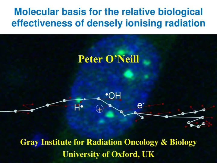
Peter ONeill OH e - H + Gray Institute for Radiation Oncology - - PowerPoint PPT Presentation

peter o neill
SMART_READER_LITE
LIVE PREVIEW

Peter ONeill OH e - H + Gray Institute for Radiation Oncology - - PowerPoint PPT Presentation

Molecular basis for the relative biological effectiveness of densely ionising radiation Peter ONeill OH e - H + Gray Institute for Radiation Oncology & Biology University of Oxford, UK Spatial distribution of events in cells


slide-1
SLIDE 1

Molecular basis for the relative biological effectiveness of densely ionising radiation

Peter O’Neill

Gray Institute for Radiation Oncology & Biology University of Oxford, UK

  • OH

e-

H●

+

slide-2
SLIDE 2
  • Track structure defines spatial distribution of

energy deposition events

  • heterogeneous and homogenous
  • Chemistry defines types of lesions and yields
  • oxygen effects
  • Sub-cellular distribution of damage defined by

ionisation density of the radiation

  • clusters of lesions (nm scale), clusters of DSB (mm scale), random

distribution

  • Distribution of DSB and clustered damage

between cells dependent on

  • the radiation dose for sparsely ionising radiation
  • the fluence for densely ionising radiations

Spatial distribution of events in cells

slide-3
SLIDE 3

Damage complexity is largely dependent on ionisation density of the radiation

30-40% low-LET = complex 90% high-LET = complex

Simple DSB Complex DSB Clustered damage

1 2 3 10 20 30 40 50 60 70 80 90 100

  • r more

percentage of total number of lesions in cluster

low LET high LET

Nikjoo, O’Neill, Goodhead, Terrissol,

  • Int. J. Radiat. Biol., 71, 467-483 (1997);

Friedland et al., Int. J. Radiat. Biol., early on line (2011)

slide-4
SLIDE 4

Maintaining Genomic Stability

DSB

Ionising Radiation Free Radicals Exogenous chemical species Replication errors

clustered DNA damage base damage SSB

+

non-homologous end joining base excision repair homologous recombination

Simple DSB Complex DSB

euchromatin heterochromatin

slide-5
SLIDE 5

Lind et al Radiation Research 160, 366-375 (2003)

Surviving fraction

dose/Gy 2 4 6 8

Co MO59K

60

Co MO59J

60

N MO59K

14 7+

N MO59J

14 7+

Co K

60

Co J

60

  • N-ions K
  • N-ions J

■ □

Co K

60

■ Deficiency in DNA damage repair: RBE of ~1

Inactivation of the major DNA DSB repair pathway, leads to similar radiosensitivity - independent of LET.

MO59J- inactive DNA-PKcs

slide-6
SLIDE 6

IR-induced DNA damage

IR

SSB DSB Base damage simple

2 or more damages within 1-2 helical turns of the DNA

Base excision repair SSB repair Non-homologous end joining Homologous recombination Single strand annealing

complex Non-DSB clustered damage

slide-7
SLIDE 7

IR-induced DSB – different types

double-ended DSB

  • prompt DSB
  • frank DSB

Non-homologous end joining

  • ne-ended DSB
  • replication induced DSB

Homologous recombination

slide-8
SLIDE 8

0.2 0.4 0.6 0.8 1 1.2 4 8 12 16 20 24 Relative Yield of DSBs Time (h)

56Fe gamma-…

a g

Double strand breaks

Variation in dynamics of DSB loss following irradiation

56Fe ion

g-radiation

gH2AX PFGE

Do differences in repair kinetics reflect DSB of different complexity? Different sub-sets of proteins recruited to some DSB?

Anderson, Harper, Cucinotta, O’Neill.

  • Radiat. Res. 174, 195-205 (2010)

Jenner, de Lara, O'Neill,Stevens,

  • Int. J. Radiat. Biol, 64, 265-273 (1993)
slide-9
SLIDE 9

6 h 24 h 0.5 h

1Gy 56Fe ions (1 GeV/nu) – DSB detection by gH2AX

Tracks of DSB (gH2AX foci) remain at longer times -

persistence of DSB reflects complexity of DSB

Jakob et al. NAR 39, 6489 (2011); Jeggo et al, EMBO J., 30, 1079 (2011)

Heterochromatin/euchromatin?

slide-10
SLIDE 10

Damage complexity- all damage substrates are not the same

Primer extension Write off

repair

Cannibalise from other car

Simple damage complex damage Very complex damage

slide-11
SLIDE 11

Dynamics of repair of DSB induced in cells irradiated at 37 ºC gH2AX as quantitative marker of DSB? Role of heterochromatin?

PFGE γ-H2AX Time (min)

60 120 180 100

% DSB remaining

50

Complex DSB and/or heterochromatin DSB

slide-12
SLIDE 12

DSB Repair in Absence of DNA-PKcs: 56Fe

10 20 30 40 50 60 70 80 90 100 4 8 12 16 20 24

Time (h)

% of cells with gamma H2AX tracks

5 10 15 20 25 30 35 40 45 50 4 8 12 16 20 24

Time (h)

% of cells with RAD 51 tracks

RAD51

gH2AX (1 Gy) (1 Gy)

M M H

Inactive DNA-PKcs Active DNA-PKcs

replication induced DSBs

late S- & G2-phase Slow Repair

Some clustered damage converted to DSBs at replication

clustered damage complex DSBs slow repair

NHEJ +DNA-PKcs

HR?

Anderson, Harper, Cucinotta, O’Neill.

  • Radiat. Res.

174, 195-205 (2010)

slide-13
SLIDE 13

Hoglund and Stenerlow, Radiation Research 155, 818-825 (2001)

small DNA fragments induced by nitrogen ions mean ~25 kbp

Does the attempted repair of DSB when clustered occur independently?

slide-14
SLIDE 14

Does loss of gH2AX foci reflect repair dynamics

  • f DSB induced by ion-particles?

multiple DSB

slide-15
SLIDE 15

RBE for DSB on LET

Co-60 gamma-rays P 250, MeV LET 0.4 He 250 MeV/u, LET 1.6 C 250 MeV/u, LET 13.8 Fe 250 MeV/u, LET 260 He 1.75 MeV/u, LET 100 C 18.33 MeV/u, LET 100 C 8.33 MeV/u, LET 201 Fe 414 MeV/u, LET 202 C 2.71 MeV/u, LET 442 Fe 115 MeV/u, LET 442

  • D. Alloni, A. Campa, M. Belli, G Esposito, L. Mariotti, M. Liotta, W. Friedland, H. Paretzke and A Ottolenghi. Radiation Research 173, 263 (2010)

Calculated number of DSB from DNA fragment distribution

slide-16
SLIDE 16

10

2

10

1

10 10

  • 1

10

  • 2

10

  • 3

20 40 60 80 100

LET (keV/um) Residual range (mm)

Mark A. Hill, Gray Institute

Variation in LET along the path of a charged particle 150 MeV Proton in water

Multiple DSB

slide-17
SLIDE 17

The effect of oxygen

dependence of OER on LET

1 10 100

LET (keV/m) OER

1 2 3

X-rays

OER generally thought to be slightly lower for carbon ions than for proton or photons. Complexity of DNA damage decreases under hypoxia

Stewart et al. Radiat. Res. 76, 587 (2011)

slide-18
SLIDE 18

2.8 2.0 1.0

OER

LET (keV/m)

1 100 10 1000 0.1

RBE

1 2 3 4 5 6 7 8

Conclusions Schematic of RBE of cell killing/OER on LET

C H

+ 6+

slide-19
SLIDE 19

Acknowledgements

Jennifer Anderson Frank Cucinotta Mark Hill Luca Mariotti