
SLIDE 1
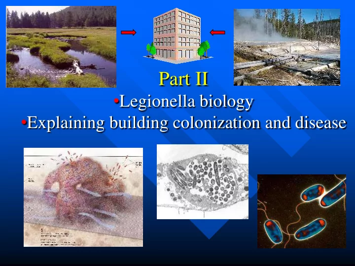
Part II
- Legionella biology
- Explaining building colonization and disease
Part II Legionella biology Explaining building colonization and - - PowerPoint PPT Presentation
Part II Legionella biology Explaining building colonization and disease Legionella are a Family of Environmental Bacteria that Live in Water Family of different species > 50 different species in genus Legionella Only half
Family of different species
Family of different species
Legionella
Other species – only in environment 90% 10%
14 serogroups Serogroup 1 (sg1)
Serogroups 2-14
85% 5%
Sub-typing, molecular fingerprinting – outbreak investigations
Surface water (lakes, streams, etc)
Actually, Legionella live in a vacuole inside of amoebae and other protozoa that live in the water
In the human lung Legionella grow in amoebae-like phagocytic cells called macrophages
Intracellular growth- amoebae and other protozoa
Some protozoa spit out these vacuoles full of Legionella
6
Surface water (lakes, streams, etc)
Thermal areas (i.e. near hot springs)
Mature biofilm
Includes amoebae feeding on these bacteria
Includes escaping amoebae and vacuoles (pellets) full of Legionella Legionella
In nature there may be hundreds of different bacteria
Sharon G. Berk, Gary Faulkner, Elizabeth Garduño, Mark
Appl Environ Microbiol. 74: 2187 (2008)
aerosols
City water (in low numbers)
Hospital-acquired LD (HA-LD)
Healthcare-associated LD
Hospital
aerosols Many different Legionella species and serogroups City water (in low numbers) drinking/aspiration from URT
Community-acquired LD (CA-LD)
Other buildings aerosols
Amost entirely L. pneumophila serogroup 1
Potable water (hot water)
Whirlpool spas Decorative fountains / Water walls
After entering from the city water in low numbers
Potable water (hot water)
Whirlpool spas Decorative fountains / Water walls
Ultrasonic humidifiers Car/bus wash
New York – 2 cases (2007) Australia – 5 cases (2008) Louisiana – 32 cases/2 deaths (1990) Sweden – 8 cases/2 deaths (2008) Wales (hotel) – 5 cases, 2 deaths (2000) Cyprus (hospital) – 11 infant cases, 2 deaths (1999)
Sources unknown or unconfirmed Susceptible person - wrong place at the wrong time aerosols
Resident alveolar macrophages
phagocytic cells – amoeba-like
Clinical presentation – not specific Chest X-ray – not specific
Clinical presentation – not specific Chest X-ray – not specific Sputum- traditional laboratory tests - negative Lack of response to antibiotics such as penicillin
May be 2-3 days post-admission
Sputum, bronchial aspirate, lung biopsy (autopsy)
Suspect colonies transferred to CYE agar with and without L-cysteine
Legionella
e.g. L.pneumophila sg1
CYE agar (selective and non-selective) With and without acid-treatment Species/serogroup antisera Sub-typing, molecular typing 5-7 days
Sputum, bronchial aspirate, lung biopsy (autopsy)
Clinical isolate
CYE agar (selective and non-selective) With and without acid-treatment Compare via sub-typing and molecular typing
Environmental isolates
Patient A Patient B
Urine antigen test - same day
Pieces of Legionella (antigens) in the blood get filtered out into the urine Results within an hour
Urine antigen test - same day
Only detects L. pneumophila sg 1 Results within an hour Will not detect all hospital-acquired Legionnaires’ disease
Resistant to penicillins and cephalosporins! Classical treatment: erythromycin or newer
New- better survival with fluoroquinolones. In
Patients still die (5-30%) due to delay in diagnosis along with the rapid progression of the disease, particularly in immunocompromised patients.