Outline Defining Sepsis Suspected infection Sepsis Definitions - - PDF document

outline defining sepsis
SMART_READER_LITE
LIVE PREVIEW

Outline Defining Sepsis Suspected infection Sepsis Definitions - - PDF document

Disclosures Shock and Sepsis 2014 Lensoo Matthew Strehlow MD FAAEM FACEP Clinical Associate Professor of EM Online educational company focused on open Co-Director Stanford Emergency Medicine International access education Stanford


slide-1
SLIDE 1

Shock and Sepsis 2014

Matthew Strehlow MD FAAEM FACEP Clinical Associate Professor of EM Co-Director Stanford Emergency Medicine International Stanford University School of Medicine

Disclosures

  • Lensoo
  • Online educational company focused on open

access education

Outline

  • Sepsis Definitions
  • Screening in the ED and prehospital
  • ProCESS
  • CVP and fluid responsiveness
  • ScvO2 and Lactate
  • Other therapies

Defining Sepsis

  • Suspected infection
  • And
  • 2 of 4 SIRS (systemic inflammatory response syndrome)
  • HR >90
  • Temp <36 or >38
  • RR >20 or PCO2 <32
  • WBC <4 or >12, >10% bands
slide-2
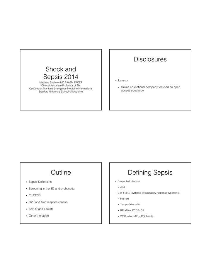
SLIDE 2

Re-defining Sepsis

  • 2001 International Sepsis Definitions Conference
  • “…the clinician goes to the bedside, identifies

myriad symptoms, and regardless of an evident infection, declares the patient to look septic.”

Re-defining Sepsis

  • 2001 International

Sepsis Definitions Conference

  • “…the clinician goes to

the bedside, identifies myriad symptoms, and regardless of an evident infection, declares the patient to look septic.”

Diagnostic criteria for sepsis

Infection, a documented or suspected, and some of the following: b General variables Fever (core temperature >38.3°C) Hypothermia (core temperature <36°C) Heart rate >90 min−1 or >2 sd above the normal value for age Tachypnea Altered mental status Significant edema or positive fluid balance (>20 mL/kg over 24 hrs) Hyperglycemia (plasma glucose >120 mg/dL or 7.7 mmol/L) in the absence of diabetesInflammatory variables Leukocytosis (WBC count >12,000 µL−1 ) Leukopenia (WBC count <4000 µL

−1 ) Normal WBC count with >10% immature forms Plasma C-

reactive protein >2 sd above the normal value Plasma procalcitonin >2 sd above the normal valueHemodynamic variables Arterial hypotension b (SBP <90 mm Hg, MAP <70, or an SBP decrease >40 mm Hg in adults or <2 sd below normal for age) So2 >70% b Cardiac index >3.5 L·min−1 ·M−23 Organ dysfunction variables Arterial hypoxemia (Pao2 /Fio2 <300) Acute

  • liguria (urine output <0.5 mL·kg−1 ·hr−1 or 45 mmol/L for at least

2 hrs) Creatinine increase >0.5 mg/dL Coagulation abnormalities (INR >1.5 or aPTT >60 secs) Ileus (absent bowel sounds) Thrombocytopenia (platelet count <100,000 µL

−1 ) Hyperbilirubinemia (plasma total bilirubin >4 mg/dL or 70

mmol/L)Tissue perfusion variables Hyperlactatemia (>1 mmol/ L) Decreased capillary refill or mottling

Severe Sepsis and Shock

  • Severe Sepsis:

Severe Sepsis: sepsis + organ dysfunction

  • Septic Shock:

Septic Shock: sepsis + hypotension after fluid resuscitation (20-30 ml/kg)

Organ System Dysfunction Respiratory Hypoxia Hematologic Low Platelets Hepatic Cardiovascular Hypotension Neurologic Renal

ProCESS Trial and EGDT

slide-3
SLIDE 3
  • Multi-center (31 USA) RCT of ED severe sepsis and

septic shock

  • Inclusion:
  • Fluid refractory septic shock OR lactate ≥4
  • Be enrolled within 2 hours of qualifying
  • Exclusion:
  • Terminal condition or DNR
  • Active severe bleeding or coagulopathy
  • Others
  • Randomized (1:1:1) to EGDT - Protocoled care -

Standard

  • EGDT 439 pts
  • Protocoled care 446 pts
  • Usual care 456 pts
slide-4
SLIDE 4

Usual Care

  • Care delivered without protocol
  • Care could not be delivered by primary site

investigator

  • At Stanford it was not delivered by ProCESS trained

physician

  • Data collected in same manner as other arms

Outcomes Randomization

  • Groups did not differ regarding baseline

characteristics or multiple severity of injury scores

slide-5
SLIDE 5

EGDT- 21% Protocol - 18.2% Usual care - 18.9%

Secondary Outcomes

  • No difference in other secondary outcomes

including

  • No difference in 90 day mortality
  • No difference in need for organ support
  • Duration of ICU/hospital stay

Care Received

  • IV fluiids
  • 3.3 L in protocol*
  • 2.8 L in EGDT*
  • 2.3 L in usual care
  • Vasopressors
  • 52.2% in protocol
  • 54.9% in EGDT
  • 44.1% in usual care*
slide-6
SLIDE 6

Care Received

  • Dobutamine
  • 1.1% in protocol
  • 8.0% in EGDT*
  • 0.9% in usual care
  • Transfusions
  • 8.3% in protocol
  • 14.4% in EGDT
  • 7.5% in usual care*

Limitations

  • Patients lower mortality than in original EGDT study. However, the most

ill 1/3 of patients showed no difference in outcomes from less ill group

  • ICU not mandated for usual care and protocol based standard therapy

groups (it was not for EGDT either but most centers cannot do CVP and ScvO2 outside of ED/ICU). This could bias in favor of a greater percentage of EGDT patients receiving ICU level care after leaving ED.

  • Fluid requirement for enrollment was altered mid-study from 20-30mL/kg

to 1 L minimum

  • Looks at patients detected early
  • Limited power to evaluate specific subgroups

ProCESS Conclusions

  • In patients that were identified rapidly and received
  • ther care such as antibiotics early there was no

benefit to protocoled care or invasive care.

ED and EMS Sepsis Screening

slide-7
SLIDE 7

Adults with Suspected Infection in the ED

Admitted = 84%

Shapiro et al. Ann Emerg Med. 2006;48:583-590.

Mortality of Patients with Suspected Infection in ED

Gille-Johnson P, et al. Scan J of Infect Dis. 2013

Mortality of Prehospital Severe Sepsis Patients

Guerra et al. JEM 2013

1 Millionth Vital Sign?

  • Shock Index = HR/SBP
  • ≥ 0.8 concerning

Berger et al. WestJEM 2013

slide-8
SLIDE 8

Summary: Sepsis Screening in the ED

  • Increasing sepsis awareness in the hospital improves
  • utcomes
  • SIRS alone is not a useful tool to screen for sepsis in the ED
  • Increasing and protocoling use of lactates and shock index

may improve sepsis identification in the ED

  • Early detection of severe sepsis and septic shock may

improve outcomes

  • Further study is needed on ED and prehospital sepsis

screening

Sepsis Management

CASE

  • 80 yo F with AMS and hypotension
  • 30 mL/kg IV NS given
  • Repeat VS
  • HR 60
  • BP 85/45
  • RR 16
  • SaO2 95% NC
slide-9
SLIDE 9

FRANK-STARLING CURVE

Goal IVF: ⬆cardiac output CO = HR x SV ∆ CO ∝ ∆ SV

Stroke Volume Preload

CONTRACTILITY EFFECTS CARDIAC OUTPUT

Stroke Volume Preload

Normal Ventricular Contractility Impaired Ventricular Contractility

CONTRACTILITY EFFECTS CARDIAC OUTPUT

Preload Stroke Volume Preload

Normal Ventricular Contractility Impaired Ventricular Contractility = small ⬆ in SV = large ⬆ in SV

FLUID RESPONSIVE

IVF bolus incr IVF bolus increases CO >10-15% eases CO >10-15%

slide-10
SLIDE 10

Marik P, P, Crit Care e Med ed 2013 “No data to support the widespread practice

  • f using CVP to guide fluid therapy”

AUC 0.56 57% of patients were fluid responders

Boyd, Crit Care e Med ed 2011 Positive fluid balance at 12 hrs and 4 days was associated with ⬆ mortality (Quartile 1 vs 4 HR 0.57, CI 0.41-0.80) Average fluid balance at 12 hours was +4.2L (day 4 +11L) CVP <8 mmHg at 12 hrs associated with lower mortality

Static measurement to determine need for fluid Dynamic measurement to determine need for fluid Measure cardiac output Measure cardiac output Give a fluid challenge

slide-11
SLIDE 11

Give a fluid challenge Give 500mL Fluid Bolus Passive Leg Raise Test

Feet at 45° 45° semi-upright

  • 200-300 mL fluid bolus
  • Lasts 30-90 seconds

Pulse Pressure Differential Stroke Volume Variability IVC Caval Index Pleth Variability Index Left Ventricular End Diastolic Area End-Expiratory Occlusion Test Doppler Monitoring

Cardiac Output

Lakhal, Ann Fren ench Anes est Rea eanima mation 2012 112 intubated ICU pts in shock received PLR ≥17% increase in SBP, then fluid responsive (+LR=26) Not helpful if increase <17% If CVP increase ≥2 mmHg then, ≥9% SBP predictive of fluid responsiveness (+LR 5.7, -LR 0.07)

slide-12
SLIDE 12

ETCO2 AS AN ESTIMATE FOR CARDIAC OUTPUT

  • ≥5% increase in ETCO2 with PLR in sedated,

mechanically ventilated patients predicts fluid responsiveness

  • +LR= 4.5-15
  • -LR= 0.1-0.3

Monnet, Intensive Care Med 2013 Garcia, Ann Intensive Care 2012

Fluids for Undifferentiated Shock

*If volume overload at any point then stop IVF and start vasopressors Undifferentiated Shock* 30 mL/kg NS or LR

YES

Observe Fluid Challenge Shock Improved?

Fluid Challenge (IVF/PLR)

Patient- MV/sedated Measure- ETCO2 Patient- CVP Available Measure- CVP & SBP Patient- No MV/CVP Measure- SBP ∆ ≥5% ∆ <5% ∆ <2 CVP & <17% SBP ∆ ≥2 CVP & <9% SBP ∆ ≥2 CVP & ≥9% SBP ∆ ≥17% SBP ∆ <17% SBP ∆ ≥17% SBP

  • Fluid Responsive
  • Indeterminate
  • Not Fluid Responsive

IV FLUIDS IN SEPSIS

  • Initial 30 mL/kg NS/LR bolus over 1 hour
  • Single CVP poor predictor of need for fluids
  • Over administration of fluids is harmful
  • Fluid challenges (PLR) and assessment of cardiac
  • utput changes are the future
slide-13
SLIDE 13
  • Which vasopressor would select?
  • A. Epinephrine
  • B. Norepinephrine
  • C. Dopamine
  • D. Vasopressin
  • Which vasopressor would select?
  • A. Epinephrine
  • B. Norepinephrine
  • C. Dopamine
  • D. Vasopressin

Vasopressors Vasopressors

  • Norepinephrine is now sole 1st line medication
  • Epinephrine is second line (swap for NE or add to

NE)

slide-14
SLIDE 14

Tissue Hypoperfusion

  • Central venous oxygen saturation (ScvO2)
  • Lactate (venous or arterial)

Tissue

ScvO2

SaO2 ScvO2 95% 75% Tissue SaO2 ScvO2 Tissue

Sepsis

ScvO2

95% 75% Tissue SaO2 ScvO2

ScvO2

Tissue 95% 55%

Sepsis

slide-15
SLIDE 15

ScvO2 Monitoring

  • Continuous reading via PreSep

catheter

  • Send serial central VBGs every

1/2 to 1 hour

Lactate Clearance, An Alternative to ScvO2

  • Check initial lactate (Time 0 hours)
  • Optimize CVP and MAP
  • Recheck lactate (Time ≥2 hours)
  • If, <10% clearance use transfusion and

dobutamine

  • Recheck, lactate every 0.5 to 1 hour

Jones A. et al. JAMA 2010;303:739-46 Jansen et al. Am J Resp Crit Care

  • Med. 2010

Tissue SaO2 ScvO2 Tissue

Sepsis

ScvO2

95% 55%

slide-16
SLIDE 16

Tissue SaO2 ScvO2 Tissue

Sepsis

ScvO2

95% 90% Shunt Tissue SaO2 ScvO2 Tis sue

Sepsis

ScvO2

95% 90%

Mortality Grouped by ScvO2

(≥90%) (<70%) (70-89%)

Pope et al. Annals of EM 2010

Markers of Possible “Occult” Hypoperfusion

  • ScvO2 <70% or

ScvO2 <70% or ≥90% 90%

  • Lactate >4 mmol/dL

Lactate >4 mmol/dL

  • Urine output <0.5ml/kg
  • Cool extremities
  • Delayed capillary refill >4 sec
  • AMS
slide-17
SLIDE 17

270 270

ScvO2 <70% or <10%⬇ lactate >3

270 Hgb <7

Transfuse Dobutamine

Hgb >10 Hgb 7-10

Signs of ischemia?

270 270 No Yes

Summary: Occult Hypoperfusion

  • ScvO2 and lactate guided therapy are options for

patients with signs of persistent hypoperfusion after fluid resuscitation and vasopressors

  • Lactate guided therapy is alternative
  • Target 10-20% reduction over 2 hours
  • Consider NTG for persistent lactate >3 (or other

markers of hypoperfusion) and normal/elevated ScvO2

Other Goals of Resuscitation Source Control

  • Indwelling lines should be

removed after alternative access has been established if it is a likely source of infection

slide-18
SLIDE 18

Steroids

  • Consider only for patients that

have sustained hypotension despite adequate fluids and vasopressors

  • No ACTH stim test

Glucose Control

  • Target <180 mg/dL
  • Consider insulin gtt if 2

consecutive values >180 mg/ dL

  • Caution with capillary blood

glucose as they may be inaccurate

Acute Lung Injury

  • Prevention
  • Keep plateau pressures ≤30 cm H2O
  • Utilize Positive end-expiratory pressure (PEEP) to

avoid alveolar collapse

  • If ARDS with PaO2/FiO2 <150 mmHg, target
  • tidal volumes 6mL/kg
  • conservative fluid strategy

Plateau pressure measures actual pressure alveoli are exposed to

Plateau Pressure

slide-19
SLIDE 19

Antimicrobial Therapy

0-2 hour broad spectrum ABX in ED Reassess in ICU Daily Recheck

Hours Hours

Summary

  • ED screening for SIRS and sepsis is not evidence

based

  • Central lines and CVP guided resuscitation remains

standard of care but US or PLR are simple early guides

  • ScvO2 is preferred over lactate guided

resuscitation but lactate guided resuscitation is a reasonable alternative