
SLIDE 1
11/ 10/ 2016 1
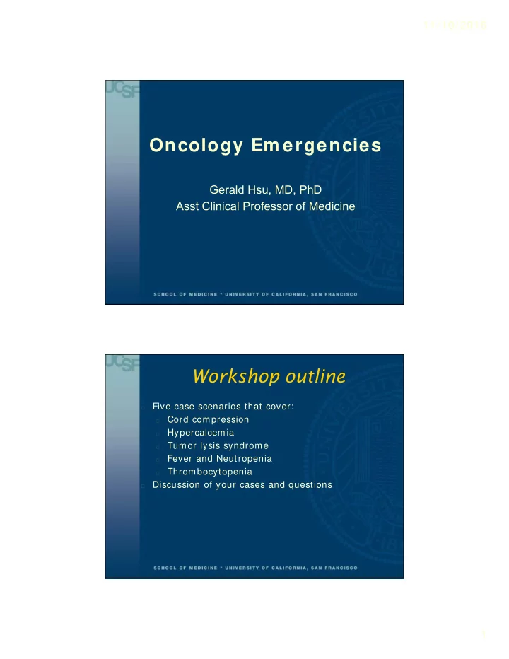
Oncology Em ergencies
Gerald Hsu, MD, PhD Asst Clinical Professor of Medicine
Workshop outline
- Five case scenarios that cover:
- Cord compression
- Hypercalcemia
- Tumor lysis syndrome
- Fever and Neutropenia
- Thrombocytopenia
- Discussion of your cases and questions