
SLIDE 1
- JOP. J Pancreas (Online) 2010 Jan 8; 11(1):72-74.
- JOP. Journal of the Pancreas - http://www.joplink.net - Vol. 11, No. 1 - January 2010. [ISSN 1590-8577]
72
CASE REPORT
Obstructive Jaundice Due to a Pancreatic Mass: A Rare Presentation of Acute Lymphoblastic Leukaemia in an Adult
Sudin Varghese Daniel1, Deven Harshad Vani2, Andrew Melvin Smith1, Quentin Antony Hill1, Krishna Viswanath Menon1
1St James’s University Hospital. Leeds, United Kingdom. 2Pinderfields General Hospital. Wakefield, United Kingdom
ABSTRACT Context To highlight a rare presentation of acute lymphoblastic leukaemia. Case report A 39-year-old man presented with a 4 month history of weight loss and a 6 week history of upper abdominal pain radiating to the back with nausea and vomiting. Liver function tests showed an obstructive picture, full blood count was normal and on computerised tomography there was diffuse enlargement of the pancreas, with dilatation of the common bile duct and intra hepatic biliary radicles. Four weeks after presenting, the white cell count became elevated with blasts on the blood film and bone marrow biopsy revealed a precursor B cell acute lymphoblastic leukaemia. After induction chemotherapy his jaundice resolved, the pancreatic mass reduced in size and he is now in a complete remission. Conclusion Acute lymphoblastic leukaemia may mimic common causes of a pancreatic mass such as adenocarcinoma and should be considered as part of the differential diagnosis when atypical features are present.
INTRODUCTION Cholestatic jaundice is an unusual presentation of acute lymphoblastic leukaemia. It is even rarer to be caused by involvement of the pancreas resulting in obstructive
- jaundice. We report a case of B cell acute
lymphoblastic leukaemia presenting as a pancreatic mass and obstructive jaundice. CASE REPORT A 39-year-old man presented with a 4-month history of 6 kg weight loss and a 6-week history of upper abdominal pain radiating to the back with nausea and
- vomiting. He had no significant past medical history
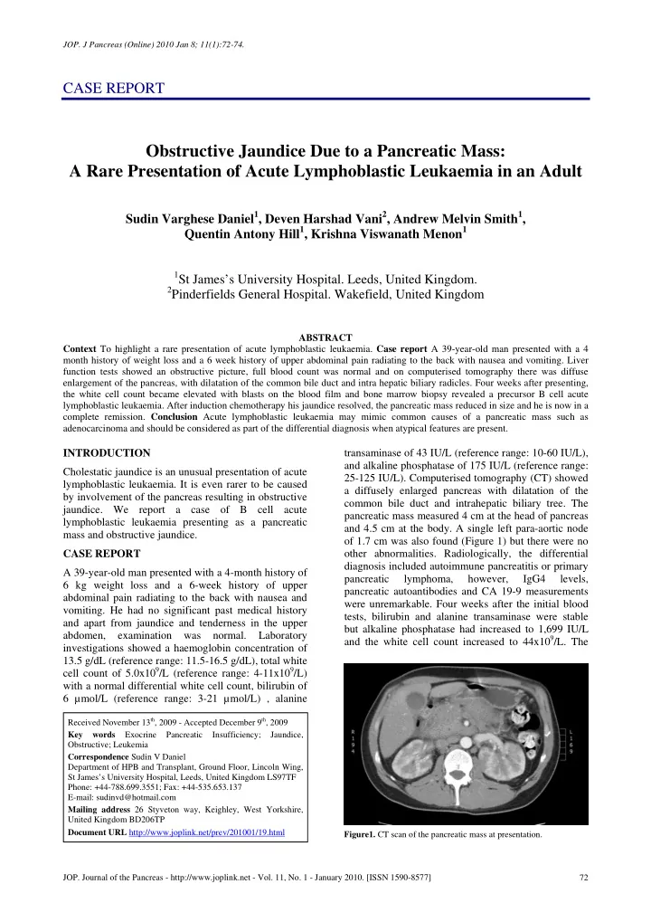
and apart from jaundice and tenderness in the upper abdomen, examination was normal. Laboratory investigations showed a haemoglobin concentration of 13.5 g/dL (reference range: 11.5-16.5 g/dL), total white cell count of 5.0x10⁹/L (reference range: 4-11x109/L) with a normal differential white cell count, bilirubin of 6 µmol/L (reference range: 3-21 µmol/L) , alanine transaminase of 43 IU/L (reference range: 10-60 IU/L), and alkaline phosphatase of 175 IU/L (reference range: 25-125 IU/L). Computerised tomography (CT) showed a diffusely enlarged pancreas with dilatation of the common bile duct and intrahepatic biliary tree. The pancreatic mass measured 4 cm at the head of pancreas and 4.5 cm at the body. A single left para-aortic node
- f 1.7 cm was also found (Figure 1) but there were no
- ther abnormalities. Radiologically, the differential
diagnosis included autoimmune pancreatitis or primary pancreatic lymphoma, however, IgG4 levels, pancreatic autoantibodies and CA 19-9 measurements were unremarkable. Four weeks after the initial blood tests, bilirubin and alanine transaminase were stable but alkaline phosphatase had increased to 1,699 IU/L and the white cell count increased to 44x109/L. The
Received November 13th, 2009 - Accepted December 9th, 2009 Key words Exocrine Pancreatic Insufficiency; Jaundice, Obstructive; Leukemia Correspondence Sudin V Daniel Department of HPB and Transplant, Ground Floor, Lincoln Wing, St James’s University Hospital, Leeds, United Kingdom LS97TF Phone: +44-788.699.3551; Fax: +44-535.653.137 E-mail: sudinvd@hotmail.com Mailing address 26 Styveton way, Keighley, West Yorkshire, United Kingdom BD206TP Document URL http://www.joplink.net/prev/201001/19.html
- Figure1. CT scan of the pancreatic mass at presentation.