Objectives The Clinical Breast Exam Revisited: Review: Whats New? - - PDF document

objectives
SMART_READER_LITE
LIVE PREVIEW

Objectives The Clinical Breast Exam Revisited: Review: Whats New? - - PDF document

Objectives The Clinical Breast Exam Revisited: Review: Whats New? Contribution of CBE to the early detection of breast cancer Cynthia Kreger, MD Variables influencing the effectiveness of the CBE Professor Clinical Internal


slide-1
SLIDE 1

1

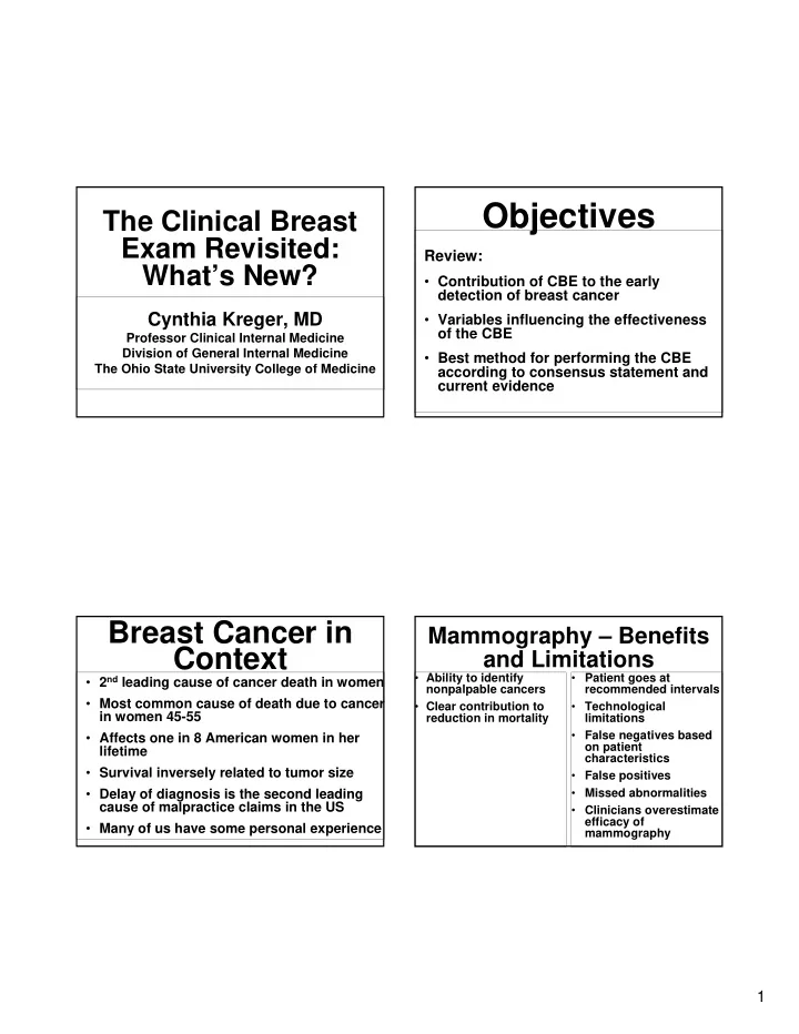
The Clinical Breast Exam Revisited: What’s New?

Cynthia Kreger, MD

Professor Clinical Internal Medicine Division of General Internal Medicine The Ohio State University College of Medicine

Review:

  • Contribution of CBE to the early

detection of breast cancer

  • Variables influencing the effectiveness
  • f the CBE
  • Best method for performing the CBE

according to consensus statement and current evidence

Objectives

Breast Cancer in Context

  • 2nd leading cause of cancer death in women
  • Most common cause of death due to cancer

in women 45-55

  • Affects one in 8 American women in her

lifetime

  • Survival inversely related to tumor size
  • Delay of diagnosis is the second leading

cause of malpractice claims in the US

  • Many of us have some personal experience

Mammography – Benefits and Limitations

  • Ability to identify

nonpalpable cancers

  • Clear contribution to

reduction in mortality

  • Patient goes at

recommended intervals

  • Technological

limitations

  • False negatives based
  • n patient

characteristics

  • False positives
  • Missed abnormalities
  • Clinicians overestimate

efficacy of mammography

slide-2
SLIDE 2

2

What is the Goal of the CBE?

Goal:

  • To detect palpable abnormalities in

asymptomatic women at an earlier stage of disease when treatment options are greater and more effective

  • To evaluate patient symptoms
  • To provide screening in women for whom

mammography is not recommended

  • To provide screening in limited resource

settings

What is the Contribution of the CBE to Early Detection?

  • Lack of RCT demonstrating CBE

reduces mortality

  • Population-based study:

71.2% of cancers identified by BSE 19.6% of cancers identified by mammogram 9.3% of cancers identified by CBE

  • Relied on recall, was in younger

women

  • More recent studies suggest that:

5.1% of malignancies detected by CBE in women with negative, benign or probably benign mammograms

  • This is over 10,000 otherwise undetected cancers per year

10.7% of cancers identified by CBE alone CBE plays a role in detection of interval cancers, in screening for women under 40, and in women who do not receive high quality mammograms or who do not follow recommendations for screening mammography

What is the Contribution of the CBE to Early Detection?

Physician Variables

  • Unconvinced about the value of the exam
  • Discomfort with the exam
  • Confidence, skill
  • Considerable variability in way the exam is taught

and performed

  • Reliance on technology to provide the answer
  • Limited time
  • Experience in detecting abnormal breast lesions

What Are The Barriers To And Variables Influencing CBE?

slide-3
SLIDE 3

3 Patient characteristics

Tissue density, nodularity, menopausal status

Tumor characteristics

Size, depth, mobility, firmness

What Are The Barriers To And Variables Influencing CBE? CBE Skills Among Graduating Primary Care Physicians

  • Only 50% examined the patient in a supine

position with arm over head

  • Only 55% performed systematic palpation
  • Only 37% examined the supraclavicular

region

  • Only 25% examined the axilla
  • Some evidence that CBE skills diminish

during training

The Components of the CBE Have Not Changed

  • Inspection
  • Nodal Evaluation
  • Breast Palpation

What’s Different Regarding Inspection?

  • Inspection

No studies document the independent benefit of inspection Taking into account limited time, inspect while palpating Increase inspection if abnormality found

  • n palpation
slide-4
SLIDE 4

4

What’s Different Regarding Inspection?

  • Inspection

Look for subtle changes such as flattening of breast contour, area of fullness, asymmetry, difference in venous pattern, scaliness of skin Findings such as erythema, retraction or dimpling, or changes in the nipple such as inversion, tend to be late signs

What’s Different Regarding Lymphatic Examination?

  • Palpation of lymph

nodes should:

  • Include the supra and

infra clavicular areas

  • Include the apical,

central, pectoral, and subscapular areas

  • Be performed with the patient seated
  • MammaCare method

Most widely studied

Recommended by CDC and the ACS

What’s Different Regarding Palpation?

  • Emphasizes the following core

competencies

Positioning Perimeter Palpation Pressure Pattern Time

What’s Different Regarding Palpation?

slide-5
SLIDE 5

5

What’s Different Regarding Positioning? The Cahan Position

Note two characteristics:

  • Position of patient’s

ipsilateral arm, the hand resting on forehead, which softens pectoralis muscle

  • Position of hips/knees to contralateral side,

which helps to distribute breast tissue centrally over chest wall

Include the Full Perimeter During Palpation

  • Perimeter as pentagon
  • Sternum to the lateral

chest wall at the mid-axillary line

  • Clavicle to below the

infra-mammary ridge

  • Junction of the shoulder with the

anterior chest, at anterior axillary line

Performing the Examination Palpation/Pressure

  • Three fingers
  • Dime-sized circles
  • Overlapping by one finger breath

with fingers sliding over breast tissue helps to ensure no areas are missed

palpate directly over nipple

Performing the Examination Palpation/Pressure

  • Pay particular attention to upper outer

quadrant, and under nipple

  • No need to assess for nipple

discharge

  • In women with breast implants -

perform the CBE in the same way

  • In women post mastectomy - palpate

all of chest wall and along incision

slide-6
SLIDE 6

6

  • Vertical strip pattern in

contrast to concentric circles

  • Palpate from distal to

proximal toward you

  • Efficacy in detecting lumps
  • Vertical strip (67.9%) vs. spoke pattern (44.7%)
  • Vertical strip (64.4%) vs. concentric circles

(38.9%)

What’s Different Regarding Pattern?

Performing the Examination Time - A Critical Variable

  • Duration of exam (and consistency of search

pattern) are the factors most consistently shown to correlate directly with sensitivity and specificity

1 minute increase in exam duration resulted in 1.8 more lumps being noted, but also increased false positive rates Optimal duration is influenced by a variety of factors: proficiency of examiner, breast size, lumpiness, body weight, tenderness A thorough exam may take up to 3-4 minutes per side

Video Demonstration

At this point I would like to share a video clip that highlights selected portions of the exam.

What Can I Take Home?

  • Don’t overestimate the efficacy of mammography, don’t

underestimate the importance of CBE

  • Use the preferred method for CBE

Include infra and supraclavicular in lyamphatic evaluation Consider the use of Cahan’s position Three level palpation, vertical strip pattern, cover full perimeter of breast tissue

  • Remember that time and consistency of search pattern

are the most critical variables

  • Any abnormality found on CBE, even in the face of a

normal mammogram, needs evaluation to appropriate resolution

slide-7
SLIDE 7

7

Adele Lipari, DO

Assistant Professor of Radiology Ohio State University Medical Center

Breast Cancer Screening and Diagnosis

Palpable mass usually ½” Mammograms detect ¼”

Craniocaudad Lateral

slide-8
SLIDE 8

8

Mammogram Paddles

  • Lowers x-ray dose
  • Reduces thickness
  • Immobilizes breast
  • Spreads out tissue

Adequate Compression

Mammograms

  • Screening
  • Diagnostic
slide-9
SLIDE 9

9

ACR Recommendations for Sreening Mammogams

  • Baseline between ages 35-40
  • Annual screening mammograms after

age 40

Screening Versus Diagnostic Mammogram

  • Screening

No breast problems No self history of breast cancer Over age 40

Diagnostic Mammogram

  • Mass
  • Persistent, pin-point pain
  • Personal history of breast Ca
  • Increase in size/firmness
  • New nipple retraction
  • Itching/flackiness of nipple
  • Spontaneous nipple discharge-

serous/bloody

slide-10
SLIDE 10

10

Duty of Referring Doctor

  • Results of Clinical Exam
  • Location of Palpable Lesion
  • Recent Needle Biopsy
  • 10% of breast cancers are

not seen by mammograms

  • r ultrasounds

Bi-Rads Code

  • Bi-Rads 1- Negative
  • Bi-Rads 2- Benign findings
  • Bi-Rads 3- Short follow up
  • Bi-Rads 4- Suggestive of Ca
  • Bi-Rads 5- Strongly suggestive
  • Bi-Rads 6- Known Ca

Ultrasound of the Breast

slide-11
SLIDE 11

11

Value of Breast US

  • Cysts
  • Margins/blood flow – solid mass
  • Lymph nodes
  • Duct evaluation
  • Silicone implant leak
  • F/U known Ca
  • Perform aspiration/biopsy

DMIST

  • Digital mammographic screening trials
  • Study to determine value of MRI and

digital mammography

slide-12
SLIDE 12

12

Digital Mammograms

  • Detect 15-28% more Ca in

premenopausal women or those

  • ver 50 with dense breasts

Acrin Guidelines for Screening MRI

  • > 20% risk of Breast Ca
  • BRCA 1 and BRCA 2 gene mutation
  • 1st degree relative with mutation
  • Strong family history
  • Chest radiation between 10-30

MRI in Contralateral Breast

  • 10% of breast ca patients develop

contralateral Ca

  • DMIST showed a 3% increase in

detection in ca patients

slide-13
SLIDE 13

13

Image Guided Biopsy

  • Stereotactic biopsy
  • Ultrasound guided biopsy

Stereotactic Breast Biopsy

Stereotactic Table

slide-14
SLIDE 14

14

Biopsy Needle

Biopsy Clip

Not Stereo Candidate

  • > 300 Pounds
  • Breast too small
  • Superficial lesion
  • Deep lesion
  • Bleeding problems
  • Unable to lie prone

Ultrasound Guided

slide-15
SLIDE 15

15

Ultrasound Biopsy

Complications of Biopsy

  • Hematoma and Infection
  • Rate = 0.1%
  • Miss Rate = 3%