
SLIDE 1
John W. Engstrom, M.D. Difficult Diangnosis - RAIN 2008 February 15, 2008
1
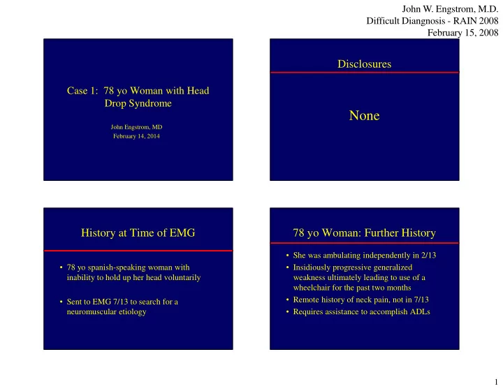
Case 1: 78 yo Woman with Head Drop Syndrome
John Engstrom, MD February 14, 2014
Disclosures
None
History at Time of EMG
- 78 yo spanish-speaking woman with
inability to hold up her head voluntarily
- Sent to EMG 7/13 to search for a
neuromuscular etiology
78 yo Woman: Further History
- She was ambulating independently in 2/13
- Insidiously progressive generalized
weakness ultimately leading to use of a wheelchair for the past two months
- Remote history of neck pain, not in 7/13
- Requires assistance to accomplish ADLs