
SLIDE 1
Nicholas Caplanis DMD MS 6/4/2012 Periodontal and Peri-Implant Considerations in Esthetic Dentistry 1 Periodontal and Peri-Implant Considerations In The Esthetic Zone
Nick Caplanis DMD MS
Private Practice Periodontics and Implant Surgery Mission Viejo, California Nick@drcaplanis.com Assistant Professor Loma Linda University
Classification of periodontal disease and conditions
- Previous classification
– 1989 world workshop
- Current classification
– 1999 international workshop
- A standard classification provides a framework for the
scientific study of disease etiology, pathogenesis and treatment as well as a standard mean of communication
Weakness of 1989 classification
- Criteria for diagnosis unclear
- Disease categories overlapped
- Too much emphasis on age of disease onset and rate of
progression which are difficult to determine
- No classification for diseases limited to gingiva
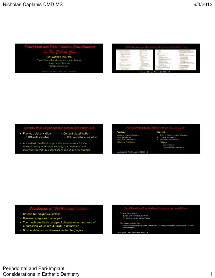
1999 Gingival and Periodontal Disease Classification
Armitage GC. Ann Periodontol 1999;4:1-6
Periodontal disease classification “Key Changes”
Previous
- No section on gingival diseases
- “Adult” Periodontitis
- “Early-onset” Periodontitis
- “Refractory” Periodontitis
Current
- Entire new section on gingival diseases
- “Chronic” Periodontitis
- “Aggressive” Periodontitis
- Additions
– Periodontal abscess – Perio-endo lesions – Acquired deformities and conditions
Armitage GC. Ann Periodontol 1999;4:1-6
Classification of periodontal disease and conditions
- Chronic periodontitis
– Typical adult onset plaque induced – Previously referred to as “adult” perio
- Aggressive periodontitis
– Previously known as pre-pubertal, juvenile perio, localized juvenile perio, rapidly progressive perio, early onset perio Armitage GC. Ann Periodontol 1999;4:1-6