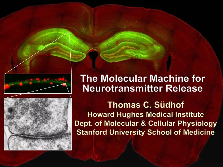
SLIDE 1
SLIDE 2
Neural Circuits Underlie Brain Function
pyramidal neurons inter- neuron interneuron
SLIDE 3 Synapses: the basic computational units
Neural Circuits Underlie Brain Function
pyramidal neurons inter- neuron interneuron
SLIDE 4 Synapses: the basic computational units
Neural Circuits Underlie Brain Function
Although synapses differ in properties, all synapses operate by the same principle
Bernard Katz - Nobel Prize, 1970
pyramidal neurons inter- neuron interneuron
SLIDE 5
An action potential invades the presynaptic nerve terminal
All Synapses Operate by the Same Principle
SLIDE 6
An action potential invades the presynaptic nerve terminal Presynaptic Ca2+-influx triggers neurotransmitter release
All Synapses Operate by the Same Principle
SLIDE 7
An action potential invades the presynaptic nerve terminal Presynaptic Ca2+-influx triggers neurotransmitter release Neurotransmitters bind to postsynaptic receptors & elicit an electrical signal
All Synapses Operate by the Same Principle
SLIDE 8
An action potential invades the presynaptic nerve terminal Presynaptic Ca2+-influx triggers neurotransmitter release Neurotransmitters bind to postsynaptic receptors & elicit an electrical signal Approach: Synaptic function is measured electrophysiologically via excitatory or inhibitory postsynaptic currents (EPSCs or IPSCs)
All Synapses Operate by the Same Principle
SLIDE 9 Synaptic transmission is rapid = 1-5 ms
- key step is neurotransmitter release
An action potential invades the presynaptic nerve terminal Presynaptic Ca2+-influx triggers neurotransmitter release Neurotransmitters bind to postsynaptic receptors & elicit an electrical signal
All Synapses Operate by the Same Principle
SLIDE 10 Synaptic transmission is rapid = 1-5 ms
- key step is neurotransmitter release
An action potential invades the presynaptic nerve terminal Presynaptic Ca2+-influx triggers neurotransmitter release Neurotransmitters bind to postsynaptic receptors & elicit an electrical signal Three basic processes enable rapid release
All Synapses Operate by the Same Principle
SLIDE 11
- 1. Synaptic vesicle fusion
Three Processes Govern Neurotransmitter Release
SLIDE 12
- 1. Synaptic vesicle fusion
- 2. Ca2+-triggering of fusion
- Very fast: ~0.1 msec
- Cooperative: ~5 Ca2+-ions
Three Processes Govern Neurotransmitter Release
SLIDE 13
- 1. Synaptic vesicle fusion
- 2. Ca2+-triggering of fusion
- Very fast: ~0.1 msec
- Cooperative: ~5 Ca2+-ions
- 3. Localized Ca2+-influx
Three Processes Govern Neurotransmitter Release
SLIDE 14
- 1. Synaptic vesicle fusion
- 2. Ca2+-triggering of fusion
- Very fast: ~0.1 msec
- Cooperative: ~5 Ca2+-ions
- 3. Localized Ca2+-influx
Three Processes Govern Neurotransmitter Release
Ca2+ Ca2+
SLIDE 15
- 1. Synaptic vesicle fusion
- 2. Ca2+-triggering of fusion
- Very fast: ~0.1 msec
- Cooperative: ~5 Ca2+-ions
- 3. Localized Ca2+-influx
Three Processes Govern Neurotransmitter Release
When I started my lab in 1986, neurotransmitter release fascinated me because of its importance, inexplicable speed, and precision – but not a single synapse component had been molecularly characterized Now – 25 years later – a molecular framework that plausibly explains release in molecular terms has emerged …
Ca2+ Ca2+
SLIDE 16
A Neurotransmitter Release Machine Mediates Fusion, Ca2+-triggering & Ca2+-Channel Tethering
SLIDE 17
A Neurotransmitter Release Machine Mediates Fusion, Ca2+-triggering & Ca2+-Channel Tethering
SLIDE 18
A Neurotransmitter Release Machine Mediates Fusion, Ca2+-triggering & Ca2+-Channel Tethering
SLIDE 19
A Neurotransmitter Release Machine Mediates Fusion, Ca2+-triggering & Ca2+-Channel Tethering
SLIDE 20
A Neurotransmitter Release Machine Mediates Fusion, Ca2+-triggering & Ca2+-Channel Tethering
Today: a brief personal account of this machine Let’s start at the beginning
SLIDE 21
- 1. Synaptic vesicle fusion
- 2. Ca2+-triggering of fusion
- Very fast: ~0.1 msec
- Cooperative: ~5 Ca2+-ions
- 3. Localized Ca2+-influx
Three Processes Govern Neurotransmitter Release
SLIDE 22
Hata et al., Nature 1993
1993 Model of Synaptic Membrane Fusion Machinery
SLIDE 23 Hata et al., Nature 1993
Based on three convergent observations:
- 1. Synaptobrevin, SNAP-25, and syntaxin are substrates for tetanus &
botulinum toxins (C. Montecucco + R. Jahn laboratories; 1992/1993)
1993 Model of Synaptic Membrane Fusion Machinery
SLIDE 24 Hata et al., Nature 1993
Based on three convergent observations:
- 1. Synaptobrevin, SNAP-25, and syntaxin are substrates for tetanus &
botulinum toxins (C. Montecucco + R. Jahn laboratories; 1992/1993)
1993 Model of Synaptic Membrane Fusion Machinery
SLIDE 25 Hata et al., Nature 1993
Based on three convergent observations:
- 1. Synaptobrevin, SNAP-25, and syntaxin are substrates for tetanus &
botulinum toxins (C. Montecucco + R. Jahn laboratories; 1992/1993)
- 2. Synaptobrevin, SNAP-25, and syntaxin form a complex, known as SNARE
complex (J. Rothman laboratory; 1993)
1993 Model of Synaptic Membrane Fusion Machinery
SLIDE 26 Hata et al., Nature 1993
Based on three convergent observations:
- 1. Synaptobrevin, SNAP-25, and syntaxin are substrates for tetanus &
botulinum toxins (C. Montecucco + R. Jahn laboratories; 1992/1993)
- 2. Synaptobrevin, SNAP-25, and syntaxin form a complex, known as SNARE
complex (J. Rothman laboratory; 1993)
1993 Model of Synaptic Membrane Fusion Machinery
SLIDE 27 Hata et al., Nature 1993
Based on three convergent observations:
- 1. Synaptobrevin, SNAP-25, and syntaxin are substrates for tetanus &
botulinum toxins (C. Montecucco + R. Jahn laboratories; 1992/1993)
- 2. Synaptobrevin, SNAP-25, and syntaxin form a complex, known as SNARE
complex (J. Rothman laboratory; 1993)
- 3. Munc18 binds to SNAREs and is homologous to Unc18 and Sec1p, proteins
known to be essential for C. elegans movements and yeast secretion by unknown mechanisms (Südhof laboratory; 1993)
1993 Model of Synaptic Membrane Fusion Machinery
SLIDE 28 Hata et al., Nature 1993
Based on three convergent observations:
- 1. Synaptobrevin, SNAP-25, and syntaxin are substrates for tetanus &
botulinum toxins (C. Montecucco + R. Jahn laboratories; 1992/1993)
- 2. Synaptobrevin, SNAP-25, and syntaxin form a complex, known as SNARE
complex (J. Rothman laboratory; 1993)
- 3. Munc18 binds to SNAREs and is homologous to Unc18 and Sec1p, proteins
known to be essential for C. elegans movements and yeast secretion by unknown mechanisms (Südhof laboratory; 1993)
1993 Model of Synaptic Membrane Fusion Machinery
SLIDE 29 Hata et al., Nature 1993
Based on three convergent observations:
- 1. Synaptobrevin, SNAP-25, and syntaxin are substrates for tetanus &
botulinum toxins (C. Montecucco + R. Jahn laboratories; 1992/1993)
- 2. Synaptobrevin, SNAP-25, and syntaxin form a complex, known as SNARE
complex (J. Rothman laboratory; 1993)
- 3. Munc18 binds to SNAREs and is homologous to Unc18 and Sec1p, proteins
known to be essential for C. elegans movements and yeast secretion by unknown mechanisms (Südhof laboratory; 1993)
Munc18 is not by-stander but central actor in membrane fusion Munc18 is absolutely essential for vesicle fusion whereas individual SNAREs are not
1993 Model of Synaptic Membrane Fusion Machinery
SLIDE 30
Verhage et al., Science 2000
Spontaneous synaptic activity in the cortex
Munc18 KO: Normal synapse formation, normal postsynaptic receptors, no presynaptic release
Munc18 KO Abolishes Synaptic Membrane Fusion
Munc18 KO
SLIDE 31
Verhage et al., Science 2000
Spontaneous synaptic activity in the cortex
Munc18 KO: Normal synapse formation, normal postsynaptic receptors, no presynaptic release A model for Munc18 function based on lots of subsequent work …
Munc18 KO Abolishes Synaptic Membrane Fusion
Munc18 KO
SLIDE 32
SNARE/SM Protein Complex Assembly Drives Fusion
SLIDE 33
SNARE/SM Protein Complex Assembly Drives Fusion
SLIDE 34
SNARE/SM Protein Complex Assembly Drives Fusion
SLIDE 35
SNARE/SM Protein Complex Assembly Drives Fusion
SLIDE 36
SNARE/SM Protein Complex Assembly Drives Fusion
SLIDE 37
SNARE/SM Protein Complex Assembly Drives Fusion
SLIDE 38
SNARE/SM Protein Complex Assembly Drives Fusion
SLIDE 39
SNARE/SM Protein Complex Assembly Drives Fusion
SLIDE 40
SNARE/SM Protein Complex Assembly Drives Fusion
SLIDE 41
SNARE/SM Protein Complex Assembly Drives Fusion
SLIDE 42
SNARE/SM Protein Complex Assembly Drives Fusion
Disease implications
SLIDE 43
SNARE/SM Protein Complex Assembly Drives Fusion
Heterozygous de novo Munc18 mutations cause Ohtahara syndrome (epileptic encephalopathy)
SLIDE 44
SNARE/SM Protein Complex Assembly Drives Fusion
Heterozygous de novo Munc18 mutations cause Ohtahara syndrome (epileptic encephalopathy)
Reinforces the importance of Munc18
SLIDE 45
SNARE/SM Protein Complex Assembly Drives Fusion
SLIDE 46 SNARE/SM Protein Complex Assembly Drives Fusion
- α-Synuclein aggregates accumulate in
Parkinson’s Disease
- Loss of α-synuclein or CSPα promotes
neurodegeneration
SLIDE 47 SNARE/SM Protein Complex Assembly Drives Fusion
- α-Synuclein aggregates accumulate in
Parkinson’s Disease
- Loss of α-synuclein or CSPα promotes
neurodegeneration
Let me illustrate …
SLIDE 48
Measured by SNARE co-immuno-precipitation
P40 P200
Burre et al., Science 2010
Deletion of Synucleins Causes Age-Dependent Impairment of SNARE-Complex Assembly
SLIDE 49
α-Synuclein overexpression in synuclein KO neurons
α-Synuclein Catalyzes SNARE-Complex Assembly
Burre et al., Science 2010
Measured as SDS-resistant SNARE complexes
SLIDE 50
α-Synuclein overexpression in synuclein KO neurons
α-Synuclein Catalyzes SNARE-Complex Assembly
Burre et al., Science 2010
Measured as SDS-resistant SNARE complexes α-Synuclein protects against some forms of neurodegeneration SNARE-complex dysfunction may contribute to Parkinson’s disease
SLIDE 51
- 1. Synaptic vesicle fusion
- 2. Ca2+-triggering of fusion
- Very fast: ~0.1 msec
- Cooperative: ~5 Ca2+-ions
- 3. Localized Ca2+-influx
Three Processes Govern Neurotransmitter Release
SLIDE 52
- 1. Synaptic vesicle fusion
- 2. Ca2+-triggering of fusion
- Very fast: ~0.1 msec
- Cooperative: ~5 Ca2+-ions
- 3. Localized Ca2+-influx
Three Processes Govern Neurotransmitter Release
At the same time as we were studying synaptic fusion, we systematically characterized synaptic vesicle proteins This approach led (among others) to the discovery of synaptotagmin, the Ca2+-sensor for neurotransmitter release
SLIDE 53
Systematic Analysis of Synaptic Vesicle Proteins Identifies Synaptotagmin-1
Südhof and Jahn, Neuron 1991
SLIDE 54
Systematic Analysis of Synaptic Vesicle Proteins Identifies Synaptotagmin-1
Südhof and Jahn, Neuron 1991
SLIDE 55
Systematic Analysis of Synaptic Vesicle Proteins Identifies Synaptotagmin-1
Ca2+-binding to Syt1 C2-domains induces lipid- and SNARE-binding
Südhof and Jahn, Neuron 1991
SLIDE 56
Systematic Analysis of Synaptic Vesicle Proteins Identifies Synaptotagmin-1
Ca2+-binding to Syt1 C2-domains induces lipid- and SNARE-binding
Südhof and Jahn, Neuron 1991
How does synaptotagmin-1 bind Ca2+, and what is its physiological significance?
SLIDE 57
C2A- Domain C2B- Domain Perin et al., Nature 1990; Brose et al., Science 1992; Davletov & Südhof, 1993; Li et al., Nature 1995; Sutton et al., Cell 1995; Chen et al., Neuron 2001
Ca2+-binding to Syt1 C2-domains induces lipid- and SNARE-binding
Y
C2A C2B
Membrane
N C
3 Ca2+ 2 Ca2+
Synaptotagmin-1 is a Synaptic Vesicle Ca2+-Binding Protein
SLIDE 58
C2A- Domain C2B- Domain
Ca2+-binding to Syt1 C2-domains induces lipid- and SNARE-binding
Y
C2A C2B
Membrane
N C
3 Ca2+ 2 Ca2+
Synaptotagmin-1 is a Synaptic Vesicle Ca2+-Binding Protein
Perin et al., Nature 1990; Brose et al., Science 1992; Davletov & Südhof, 1993; Li et al., Nature 1995; Sutton et al., Cell 1995; Chen et al., Neuron 2001
SLIDE 59
C2A- Domain C2B- Domain
Ca2+-binding to Syt1 C2-domains induces lipid- and SNARE-binding
Y
C2A C2B
Membrane
N C
3 Ca2+ 2 Ca2+
Architecture of Synaptotagmin-1 Ca2+-Binding Sites
Perin et al., Nature 1990; Brose et al., Science 1992; Davletov & Südhof, 1993; Li et al., Nature 1995; Sutton et al., Cell 1995; Chen et al., Neuron 2001
SLIDE 60
C2A- Domain C2B- Domain
Ca2+-binding to Syt1 C2-domains induces lipid- and SNARE-binding
Y
C2A C2B
Membrane
N C
3 Ca2+ 2 Ca2+
Architecture of Synaptotagmin-1 Ca2+-Binding Sites
Perin et al., Nature 1990; Brose et al., Science 1992; Davletov & Südhof, 1993; Li et al., Nature 1995; Sutton et al., Cell 1995; Chen et al., Neuron 2001
Does knockout of Syt1 impair Ca2+-triggered release?
SLIDE 61
Release stimulated by isolated action potentials
Wild-type Syt1 KO
Synaptotagmin-1 is Essential for Ca2+-Triggered Neurotransmitter Release
Geppert et al., Cell 1994
Fast Ca2+-triggered release is gone …
SLIDE 62 Hypertonic sucrose stimulates release by a Ca2+- independent mechanism
Wildtype Syt1 KO
+ Sucrose Synaptotagmin is ONLY required for Ca2+-triggered fusion
Synaptotagmin-1 is Not Essential for Sucrose-Stimulated Neurotransmitter Release
Geppert et al., Cell 1994
SLIDE 63 Y
C2A C2B
Membrane
N C
3 Ca2+ 2 Ca2+
- Synaptotagmin-1 is a synaptic vesicle Ca2+-binding protein
- Synaptotagmin-1 is essential for fast Ca2+-triggered release
However, synaptotagmin does not act alone - it needs an accomplice = complexin Synaptotagmin-1 is a Synaptic Vesicle Ca2+-Sensor Essential for Ca2+-Triggered Vesicle Fusion
SLIDE 64
A Neurotransmitter Release Machine Mediates Fusion, Ca2+-triggering & Ca2+-Channel Tethering
McMahon et al., Cell 1995; Chen et al., Neuron 2002
SLIDE 65
A Neurotransmitter Release Machine Mediates Fusion, Ca2+-triggering & Ca2+-Channel Tethering
McMahon et al., Cell 1995; Chen et al., Neuron 2002
SLIDE 66
A Neurotransmitter Release Machine Mediates Fusion, Ca2+-triggering & Ca2+-Channel Tethering
McMahon et al., Cell 1995; Chen et al., Neuron 2002
Complexin is an essential activator of synaptotagmin-1 that is evolutionarily conserved – an example
SLIDE 67
Nematostella vectensis (cnideria) Encodes synapto- tagmins & complexins
Nomastella Complexin Functions in Mouse Neurons
Yang et al., unpublished
SLIDE 68
Nematostella vectensis (cnideria) Encodes synapto- tagmins & complexins complexin
Mus Mus musc muscul ulus us
Nomastella Complexin Functions in Mouse Neurons
Yang et al., unpublished
SLIDE 69
Nematostella vectensis (cnideria) Encodes synapto- tagmins & complexins EPSCs complexin
Mus Mus musc muscul ulus us
Nomastella Complexin Functions in Mouse Neurons
Yang et al., unpublished
SLIDE 70 Y
C2A C2B
Membrane
N C
3 Ca2+ 2 Ca2+
- Synaptotagmin-1 is a synaptic vesicle Ca2+-binding protein
- Synaptotagmin-1 is essential for fast Ca2+-triggered release
- Synaptotagmin-1 uses complexin as essential co-activator
Synaptotagmin-1 is a Synaptic Vesicle Ca2+-Sensor Essential for Ca2+-Triggered Vesicle Fusion This is where we stood in 1995
SLIDE 71
Südhof Laboratory ~1995
SLIDE 72 We had – together with others – identified the major components
- f the synaptic vesicle membrane fusion machinery and
described a candidate Ca2+-sensor for fusion
Südhof Laboratory ~1995
SLIDE 73 We had – together with others – identified the major components
- f the synaptic vesicle membrane fusion machinery and
described a candidate Ca2+-sensor for fusion
Südhof Laboratory ~1995
Yutaka Hata
SLIDE 74 We had – together with others – identified the major components
- f the synaptic vesicle membrane fusion machinery and
described a candidate Ca2+-sensor for fusion
Südhof Laboratory ~1995
Yutaka Hata Martin Geppert
SLIDE 75 We had – together with others – identified the major components
- f the synaptic vesicle membrane fusion machinery and
described a candidate Ca2+-sensor for fusion
Südhof Laboratory ~1995
Yutaka Hata Harvey McMahon Martin Geppert
SLIDE 76 We had – together with others – identified the major components
- f the synaptic vesicle membrane fusion machinery and
described a candidate Ca2+-sensor for fusion
Südhof Laboratory ~1995
Yutaka Hata Harvey McMahon Martin Geppert Cai Li
SLIDE 77 Südhof Laboratory ~1995
Yutaka Hata Harvey McMahon Martin Geppert Cai Li not shown: Mark Perin, Nils Brose, Bazbek Davletov
We had – together with others – identified the major components
- f the synaptic vesicle membrane fusion machinery and
described a candidate Ca2+-sensor for fusion HOWEVER: Many doubted that SNARE & SM proteins ‘do’ membrane fusion, others suggested that synaptotagmin is a scaffold but NOT a Ca2+-sensor for fusion, and we had no idea how Ca2+-influx is localized to the site of vesicle fusion
SLIDE 78
Remainder of the talk: How we addressed the issues of Ca2+-triggering of fusion and of Ca2+-influx Major question: Does Ca2+-binding to synaptotagmin-1 really trigger fast release?
SLIDE 79
C2A- Domain C2B- Domain
Ca2+-binding to Syt1 C2-domains induces lipid- and SNARE-binding
Y
C2A C2B
Membrane
N C
3 Ca2+ 2 Ca2+
Perin et al., Nature 1990; Brose et al., Science 1992; Davletov & Südhof, 1993; Li et al., Nature 1995; Sutton et al., Cell 1995; Chen et al., Neuron 2001
Architecture of Synaptotagmin-1 Ca2+-Binding Sites
SLIDE 80
C2A- Domain C2B- Domain
Ca2+-binding to Syt1 C2-domains induces lipid- and SNARE-binding
Y
C2A C2B
Membrane
N C
3 Ca2+ 2 Ca2+
Perin et al., Nature 1990; Brose et al., Science 1992; Davletov & Südhof, 1993; Li et al., Nature 1995; Sutton et al., Cell 1995; Chen et al., Neuron 2001
Design mutations that shift the Ca2+-affinity of synaptotagmin-1 during SNARE- or phospholipid binding
Architecture of Synaptotagmin-1 Ca2+-Binding Sites
SLIDE 81
C2A- Domain C2B- Domain
Ca2+-binding to Syt1 C2-domains induces lipid- and SNARE-binding
Y
C2A C2B
Membrane
N C
3 Ca2+ 2 Ca2+
Fernandez-Chacon et al., Nature 2001; Pang et al., J. Neurosci. 2006 D232N R233Q
Design mutations that shift the Ca2+-affinity of synaptotagmin-1 during SNARE- or phospholipid binding
Architecture of Synaptotagmin-1 Ca2+-Binding Sites
SLIDE 82
R233Q mutant Syt1
Ca2+-dependent binding of native brain Syt1 to liposomes Ca2+-dependent co-IP of native brain Syt1 with SNARE complexes
D232N mutant Syt1
D232N WT
Adjacent C2A-Domain Mutations (D232N & R233N) Differentially Alter Synaptotagmin-1 Ca2+-Affinity
Fernandez-Chacon et al., Nature 2001; Pang et al., J. Neurosci. 2006
Effect on Ca2+-triggered neurotransmitter release?
SLIDE 83
C2A C2B
Membrane
Y
N C
3 Ca2+ 2 Ca2+ Ca2+-titration of release
R233Q decreases Ca2+-affinity during phospholipid binding
Synaptotagmin-1 is a Ca2+-Sensor for Synaptic Vesicle Fusion
D232N increases Ca2+-dependent SNARE binding
Fernandez-Chacon et al., Nature 2001; Pang et al., J. Neurosci. 2006
Formally proved that Ca2+-binding to synaptotagmin-1 triggers neurotransmitter release
SLIDE 84 Y
C2A C2B
Membrane
N C
3 Ca2+ 2 Ca2+
- Synaptotagmin-1 is a synaptic vesicle Ca2+-binding protein
- Synaptotagmin-1 is essential for fast Ca2+-triggered release
- Synaptotagmin-1 uses complexin as essential co-activator
- Ca2+-binding to Synaptotagmin-1 triggers fast release
However, mammals express 16 synaptotagmins! Synaptotagmin-1 is a Ca2+-Sensor for Synaptic Vesicle Fusion
SLIDE 85
C2A C2B
Membrane
Y
N C
3 Ca2+ 2 Ca2+
C2A C2B N C
3 Ca2+ 2 Ca2+
C2A C2B N C
3 Ca2+ 2 Ca2+ Membrane
S S Syt1, Syt2, Syt7, and Syt9 Syt3, Syt5, Syt6, and Syt10
Eight other synaptotagmins do not bind Ca2+ Two Classes of Synaptotagmins Bind Ca2+
Which synaptotagmins are Ca2+-sensors for fast release?
SLIDE 86
Xu et al., Neuron 2007
Rescue of Syt1 KO neurons
Syt1, Syt2, and Syt9 Rescue Syt1 KO Phenotype
SLIDE 87
Xu et al., Neuron 2007
Rescue of Syt1 KO neurons
Syt1, Syt2, and Syt9 Rescue Syt1 KO Phenotype
Syt1, Syt2, and Syt9 selectively rescue fast release in Syt1 KO neurons, but with distinct properties – whereas Syt7 does NOT rescue
SLIDE 88 C2A C2B
Membrane
Y
N C
3 Ca2+ 2 Ca2+
C2A C2B N C
3 Ca2+ 2 Ca2+
C2A C2B N C
3 Ca2+ 2 Ca2+ Membrane
S S Syt1, Syt2, Syt7, and Syt9 Syt3, Syt5, Syt6, and Syt10
Two Classes of Synaptotagmins Bind Ca2+
Two new issues:
- 1. Why does the Syt1 KO have a phenotype if Syt2 and
Syt9 can compensate?
- 2. Why doesn’t Syt7 function in release if it is so similar to
- ther ‘blue’ synaptotagmins?
SLIDE 89
Quantitation of Synaptotagmin mRNA Levels in Single Hippocampal Neurons: Syt2 and Syt9 are Absent
Bacaj et al., Neuron 2013 each column = 1 neuron
SLIDE 90
Bacaj et al., Neuron 2013 each column = 1 neuron
Syt2 & Syt9 are not expressed but Syt7 is highly expressed What does Syt7 do? Recall the initial KO results …
Quantitation of Synaptotagmin mRNA Levels in Single Hippocampal Neurons: Syt2 and Syt9 are Absent
SLIDE 91
Release stimulated by isolated action potentials
Wild-type Syt1 KO
Synaptotagmin-1 is Essential for Ca2+-Triggered Neurotransmitter Release
Geppert et al., Cell 1994
Some residual Ca2+-triggered release remains in synaptotagmin-1 KO neurons
SLIDE 92
Synaptotagmin-7 Deletion Impairs Remaining Ca2+-Triggered Release in Syt1 KO Neurons
Bacaj et al., Neuron 2013
SLIDE 93
C2A C2B
Membrane
Y
N C
3 Ca2+ 2 Ca2+
C2A C2B N C
3 Ca2+ 2 Ca2+
C2A C2B N C
3 Ca2+ 2 Ca2+ Membrane
S S Syt1, Syt2, Syt7, and Syt9 Syt3, Syt5, Syt6, and Syt10
Two Classes of Synaptotagmins Bind Ca2+
All blue synaptotagmins function in synaptic vesicle fusion but exhibit different Ca2+-triggering kinetics – function also in neuroendecrine/hormone secretion, mast cell degranulation etc.
What about red synaptotagmins? Focus on Syt10 …
SLIDE 94 Double immuno
scence labeling
Cao et al., Cell 2011
Synaptotagmin-10 Co-Localizes with IGF-1 in Olfactory Bulb Neurons
SLIDE 95
Synaptotagmin-10 Knockout Impairs Depolarization-Induced IGF-1 Secretion IGF-1 Secretion
Syt1 KD control Conditional Syt10 KO
Syt10 KO Syt10 KO
Cao et al., Cell 2011
Loss of IGF-1 secretion decreases neuron size and synapse numbers – rescue with IGF-1 Syt1 KD control
SLIDE 96 IPSCs Capacitance Syt10 is a Ca2+-sensor for IGF-1 exocytosis – does Syt10 use complexin as a co-factor?
Syt10 KO Decreases Total Synaptic Responses & Capacitance of Neurons - Rescue with IGF-1
Syt10 KO Syt10 KO Syt10 KO Syt10 KO Syt10 KO Syt10 KO
Cao et al., Cell 2011
SLIDE 97
Cao et al., J. Neurosci. 2012
IGF-1 secretion measured at different extracellular Ca2+- concentrations
Complexin Depletion Impairs Synaptotagmin-10 Dependent IGF-1 Secretion
Implications for synaptotagmin function
SLIDE 98
Multiple Pathways of Ca2+-Triggered Exocytosis Controlled by Different Synaptotagmins
Diverse non-redundant synaptotagmins use the same complexin-dependent mechanism for different Ca2+-dependent membrane fusion reactions
SLIDE 99 Y
C2A C2B
Membrane
N C
3 Ca2+ 2 Ca2+
- Synaptotagmin-1 is a synaptic vesicle Ca2+-binding protein
- Synaptotagmin-1 is essential for fast Ca2+-triggered release
- Synaptotagmin-1 uses complexin as essential co-activator
- Ca2+-binding to Synaptotagmin-1 triggers fast release
- Other synaptotagmins perform analogous functions in
Ca2+-triggered release with complexin as co-factor
Synaptotagmins Are Universal Ca2+-Sensors for Ca2+-Triggered Vesicle Fusion
SLIDE 100
- 1. Synaptic vesicle fusion
- 2. Ca2+-triggering of fusion
- Very fast: ~0.1 msec
- Cooperative: ~5 Ca2+-ions
- 3. Localized Ca2+-influx
Three Processes Govern Neurotransmitter Release
These studies thus established synaptotagmins as Ca2+-sensors for membrane fusion and generalized their functions in most if not all Ca2+-dependent fusion reactions What about Ca2+-influx?
SLIDE 101
- 1. Synaptic vesicle fusion
- 2. Ca2+-triggering of fusion
- Very fast: ~0.1 msec
- Cooperative: ~5 Ca2+-ions
- 3. Localized Ca2+-influx
Three Processes Govern Neurotransmitter Release
SLIDE 102
- 1. Synaptic vesicle fusion
- 2. Ca2+-triggering of fusion
- Very fast: ~0.1 msec
- Cooperative: ~5 Ca2+-ions
- 3. Localized Ca2+-influx
Three Processes Govern Neurotransmitter Release
Ca2+ Ca2+
SLIDE 103
- 1. Synaptic vesicle fusion
- 2. Ca2+-triggering of fusion
- Very fast: ~0.1 msec
- Cooperative: ~5 Ca2+-ions
- 3. Localized Ca2+-influx
Three Processes Govern Neurotransmitter Release
Without localized Ca2+-influx at the active zone, action potentials and release become uncoupled, and release is desynchronized and decelerated
Ca2+ Ca2+
The importance of localized Ca2+-influx cannot be
- verestimated – like in real estate, location is everything!
SLIDE 104
A Neurotransmitter Release Machine Mediates Fusion, Ca2+-triggering & Ca2+-Channel Tethering
SLIDE 105
RIM is the central component of the active zone
A Neurotransmitter Release Machine Mediates Fusion, Ca2+-triggering & Ca2+-Channel Tethering
SLIDE 106
Kaeser et al., Cell 2011; Deng et al., Neuron 2011; Han et al., Neuron 2011
Control RIM KO (cDKO)
EPSCs IPSCs Is release impaired because of a defect in Ca2+-influx?
Deletion of RIM Severely Impairs Release
SLIDE 107
Speed & synchronicity of IPSCs
Kaeser et al., Cell 2011; Deng et al., Neuron 2011; Han et al., Neuron 2011
RIM Deletion Decelerates & Desynchronizes Release: Renders Release Sensitive to Slow Ca2+-Buffers
SLIDE 108
Speed & synchronicity of IPSCs
Kaeser et al., Cell 2011; Deng et al., Neuron 2011; Han et al., Neuron 2011
Effect of slow Ca2+-buffers
Consistent with impaired Ca2+-influx measure the role of RIM in Ca2+-influx directly
RIM Deletion Decelerates & Desynchronizes Release: Renders Release Sensitive to Slow Ca2+-Buffers
SLIDE 109
Pipette
dendrites Neuron filled with: Tracer = Alexa594 + Ca2+-indicator = Fluo5F axon with presynaptic terminals
Measurement of Ca2+-Transients in Hippocampal Neurons
Kaeser et al., Cell 2011
SLIDE 110
Pipette
dendrites Neuron filled with: Tracer = Alexa594 + Ca2+-indicator = Fluo5F axon with presynaptic terminals Ca2+- signal action potential Kaeser et al., Cell 2011
Measurement of Ca2+-Transients in Hippocampal Neurons
SLIDE 111
Control RIM KO
RIM Deletion Impairs Presynaptic Ca2+-Influx
+
Rescue Presynaptic Terminal
Kaeser et al., Cell 2011
SLIDE 112
Control RIM KO
+
Rescue Dendrites
RIM Deletion Impairs Presynaptic Ca2+-Influx
Presynaptic Terminal
Kaeser et al., Cell 2011
SLIDE 113
A Neurotransmitter Release Machine Mediates Fusion, Ca2+-triggering & Ca2+-Channel Tethering
RIM also mediates synaptic vesicle docking, enable synaptic plasticity, and activates Munc13 for vesicle priming
SLIDE 114
- 1. Synaptic vesicle fusion
- 2. Ca2+-triggering of fusion
- Very fast: ~0.1 msec
- Cooperative: ~5 Ca2+-ions
- 3. Localized Ca2+-influx
Three Processes Govern Neurotransmitter Release
SLIDE 115
A Neurotransmitter Release Machine Mediates Fusion, Ca2+-triggering & Ca2+-Channel Tethering
SLIDE 116
- 1. Synaptic vesicle fusion
- 2. Ca2+-triggering of fusion
- Very fast: ~0.1 msec
- Cooperative: ~5 Ca2+-ions
- 3. Localized Ca2+-influx
Three Processes Govern Neurotransmitter Release
SLIDE 117
A Neurotransmitter Release Machine Mediates Fusion, Ca2+-triggering & Ca2+-Channel Tethering
SLIDE 118
- 1. Synaptic vesicle fusion
- 2. Ca2+-triggering of fusion
- Very fast: ~0.1 msec
- Cooperative: ~5 Ca2+-ions
- 3. Localized Ca2+-influx
Three Processes Govern Neurotransmitter Release
Ca2+ Ca2+
SLIDE 119
A Neurotransmitter Release Machine Mediates Fusion, Ca2+-triggering & Ca2+-Channel Tethering
Functionally, the fusion, Ca2+-triggering, and active zone complexes form a single interacting nanomachine mediating fast transmitter release
SLIDE 120
Reinhard Jahn Jose Rizo Robert E. Hammer Rob Malenka Jianyuan Sun Xinran Liu Axel Brunger Ralf Schneggenburger Chuck Stevens Jie Shen Jakob Sorensen
Mark Perin Martin Geppert Harvey McMahon Cai Li Nils Brose Ok-Ho Shin Sreeganga Chandra Anton Maximov Jun Xu Matthijs Verhage Zhiping Pang Weiping Han Lunbin Deng Xiaofei Yang Claudio Acuna Dick Wu Taulant Bacaj Peng Cao Peng Zhou Yea-Jin Kaeser-Woo Manu Sharma Jacqueline Burre Rafael Fernandez-Chacon Pascal Kaeser Victor P. Whittaker Michael S. Brown Joseph L. Goldstein
Key Mentors Key Students & Postdocs Key Collaborators
Funding Sources
UT Southwestern Medical School Stanford University School of Medicine
Key Institutions