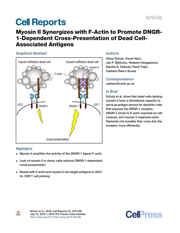

Article Myosin II Synergizes with F-Actin to Promote DNGR- 1-Dependent Cross-Presentation of Dead Cell- Associated Antigens Graphical Abstract Authors Oliver Schulz, Pavel Han � c, Jan P. Bo ¨ ttcher, Robbert Hoogeboom, Sandra S. Diebold, Pavel Tolar, Caetano Reis e Sousa Correspondence caetano@crick.ac.uk In Brief Schulz et al. show that dead cells lacking myosin II have a diminished capacity to serve as antigen donors for dendritic cells that express the DNGR-1 receptor. DNGR-1 binds to F-actin exposed on cell corpses, and myosin II organizes actin filaments into bundles that cross-link the receptor more efficiently. Highlights d Myosin II amplifies the activity of the DNGR-1 ligand F-actin d Lack of myosin II in donor cells reduces DNGR-1-dependent cross-presentation d Beads with F-actin and myosin II can target antigens to cDC1 for CD8 T cell priming Schulz et al., 2018, Cell Reports 24 , 419–428 July 10, 2018 ª 2018 The Francis Crick Institute. https://doi.org/10.1016/j.celrep.2018.06.038
Cell Reports Article Myosin II Synergizes with F-Actin to Promote DNGR-1-Dependent Cross-Presentation of Dead Cell-Associated Antigens Oliver Schulz, 1 Pavel Han � c, 1,5 Jan P. Bo ¨ ttcher, 1 Robbert Hoogeboom, 2,6 Sandra S. Diebold, 3 Pavel Tolar, 2,4 and Caetano Reis e Sousa 1,7, * 1 Immunobiology Laboratory, The Francis Crick Institute, 1 Midland Road, London NW1 1AT, UK 2 Immune Receptor Activation Laboratory, The Francis Crick Institute, 1 Midland Road, London NW1 1AT, UK 3 Biotherapeutics Division, National Institute for Biological Standards and Control, Potters Bar, Hertfordshire EN6 3QG, UK 4 Division of Immunology and Inflammation, Imperial College London, Du Cane Road, London SW7 2AZ, UK 5 Present address: Department of Microbiology and Immunobiology, Harvard Medical School, Boston, MA 02115, USA 6 Present address: Department of Haemato-Oncology, Faculty of Life Sciences and Medicine, King’s College London, London SE5 9NU, UK 7 Lead Contact *Correspondence: caetano@crick.ac.uk https://doi.org/10.1016/j.celrep.2018.06.038 SUMMARY within healthy cells but are released or exposed by dead cells following loss of plasma membrane integrity. Recognition of Conventional type 1 DCs (cDC1s) excel at cross- DAMPs by innate immune receptors, including C-type lectin re- presentation of dead cell-associated antigens partly ceptors (CLRs) on myeloid cells, promotes production of pro- inflammatory mediators (Chen and Nun ˜ ez, 2010; Rock et al., because they express DNGR-1, a receptor that 2011; Sancho and Reis e Sousa, 2013; Zelenay and Reis e recognizes exposed actin filaments on dead cells. Sousa, 2013). In dendritic cells (DCs), DAMP recognition also In vitro polymerized F-actin can be used as a syn- plays a role in the extraction of dead cell-associated antigens thetic ligand for DNGR-1. However, cellular F-actin for presentation to CD8 + T cells in a process called cross-pre- is decorated with actin-binding proteins, which sentation (Cruz et al., 2017; Han � c et al., 2016a; Joffre et al., could affect DNGR-1 recognition. Here, we demon- 2012). The most efficient cross-presenting DCs in the mouse, strate that myosin II, an F-actin-associated motor initially identified by the expression of CD8 a in lymphoid organs protein, greatly potentiates the binding of DNGR-1 and CD103 in peripheral tissues (Joffre et al., 2012; Shortman to F-actin. Latex beads coated with F-actin and and Heath, 2010), have been renamed conventional type 1 myosin II are taken up by DNGR-1 + cDC1s, and DCs (cDC1s) (Guilliams et al., 2014; Schraml and Reis e Sousa, 2015) and require the transcription factors Batf3 (Hildner et al., antigen associated with those beads is efficiently cross-presented to CD8 + T cells. Myosin II-deficient 2008), ID2 (Hacker et al., 2003), and IRF8 (Aliberti et al., 2003; Schiavoni et al., 2002) for their development. These cells are necrotic cells are impaired in their ability to stimu- also found in humans and can be identified across species by late DNGR-1 or to serve as substrates for cDC1 cross-presentation to CD8 + T cells. These results expression of the chemokine receptor XCR1 (Dorner et al., 2009), as well as high levels of the CLR DNGR-1 (also known provide insights into the nature of the DNGR-1 as CLEC9A) (Caminschi et al., 2008; Huysamen et al., 2008; ligand and have implications for understanding im- Poulin et al., 2012, 2010; Sancho et al., 2008). In addition to mune responses to cell-associated antigens and acting as a cDC1 marker, DNGR-1 is a DAMP receptor. It is ex- for vaccine design. pressed as a dimeric transmembrane protein with two C-type lectin-like domains (CTLDs) that face the extracellular space INTRODUCTION (or the lumen of endosomes) and bind to the cytoskeletal component F-actin, which is exposed in dead cells (Ahrens Cell death resulting from infection or tissue injury can lead et al., 2012; Zhang et al., 2012). DNGR-1 binding to F-actin on to release of damage-associated molecular patterns (DAMPs) cell corpses encountered or ingested by cDC1 provokes (Land, 2003) with immunomodulatory activity (Chen and Nun ˜ ez, signaling via Syk and somehow allows endocytic cargo to be 2010; Land et al., 1994; Matzinger, 1994; Rock et al., 2011). shuttled into the cross-presentation pathway (Zelenay et al., DAMPs are pre-synthesized cellular molecules such as metab- 2012). Consistent with a key role for DNGR-1 and cDC1 in cross-presentation of dead cell-associated antigens, loss of olites (ATP and uric acid), nucleic acids (DNA and RNA), or pro- DNGR-1 in mice reduces cross-priming of CD8 + cytotoxic teins (heat shock proteins [HSPs] and HMGB1) but can also be considered to include mediators actively produced by the dying T cells against model antigens contained within necrotic cells cell as a result of death receptor pathways intersecting with nu- and of viral antigens expressed by cells infected with cytopathic clear factor k B (NF- k B) signaling (Yatim et al., 2015; Zelenay viruses (Iborra et al., 2012; Sancho et al., 2009; Zelenay et al., and Reis e Sousa, 2013). DAMPs are normally sequestered 2012). Cell Reports 24 , 419–428, July 10, 2018 ª 2018 The Francis Crick Institute. 419 This is an open access article under the CC BY license (http://creativecommons.org/licenses/by/4.0/).
Figure 1. Addition of Myosin II to F-Actin Promotes DNGR-1 Binding Serial (2-fold) dilutions from top to bottom (black A B wedge) of in vitro polymerized F-actin (top con- centration: 0.4 m M in E and 0.2 m M in B–D and G) or F-actin complexed with myosin II (top concentra- tion: 0.04 m M in E and 0.2 m M in G) were analyzed by dot blot. Arrows indicate PBS control dots. (A) Schematic representation of soluble DNGR-1 reagents: ECD dimer (left) and CTLD monomer (right). (B) DNGR-1 ECD and DNGR-1 CTLD (20 m g/mL ) binding to immobilized F-actin and HeLa cell lysate. (C and D) Pre-incubation of immobilized F-actin with either blocking buffer (control), myosin II or a -actinin (C) or blocking buffer (control), spectrin or tropomyosin/troponin (D) (all at 10 m g/ml). (E and F) Titration of F-actin and F-actin and myosin II complexes (the latter starts at 10-fold lower concentration) (E) and dose-response curve C D E (F) after quantitation of the signal in (E) using ImageJ software. (G) DNGR-1 ECD and DNGR-1 CTLD (20 m g/mL) binding to immobilized F-actin and F-actin and myosin II. Data are representative of 2 (C, D, and G) and 3 (B, E, and F) independent experiments. eration of contractile force, and actin- dependent cell motility (dos Remedios et al., 2003; Pollard and Cooper, 1986). This prompted us to test purified ABPs for the ability to potentiate or block DNGR-1 binding to F-actin. Here, we report that most ABPs tested did not F G affect the interaction of DNGR-1 with F-actin, with the exception of the motor protein myosin II. F-actin combined with myosin II bound to DNGR-1 more effi- ciently than naked F-actin and showed increased agonistic activity. Consistent with that notion, antigen particles bearing F-actin and myosin II were efficiently taken up and cross-presented by DNGR-1 + cDC1s, while corpses of cells lacking myosin II were reduced in their ability to stimulate DNGR-1 and to serve as antigen sources for cross-presenta- tion. Our results indicate that F-actin We have solved the structure of DNGR-1 in complex with sin- and myosin complexes are the physiological substrates for gle-actin filaments using cryo-electron microscopy (Han � c et al., DNGR-1-dependent recognition of dead cells and can be ex- 2015). The structure showed that the CTLDs of DNGR-1 bind to ploited for the purpose of vaccination. the interface of the two F-actin protofilaments and confirms that naked F-actin is sufficient as a DNGR-1 ligand (Han � c et al., RESULTS 2015). However, in the context of dead cell recognition, engage- ment of DNGR-1 could involve an additional factor or factors. Myosin II Increases the Binding of DNGR-1 to F-Actin This is because F-actin in cells is always coated with actin-bind- DNGR-1 binding to ligand can be monitored experimentally by ing proteins (ABPs), a group of more than 200 proteins that asso- using the soluble extracellular domain (ECD) of the DNGR-1 ciate with G- and/or F-actin and regulate its polymerization, gen- dimeric receptor or the monomeric CTLD (Figure 1A) (Ahrens Cell Reports 24 , 419–428, July 10, 2018 420
Recommend
More recommend