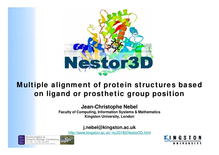

Multiple alignment of protein structures based on ligand or prosthetic group position Jean-Christophe Nebel Faculty of Computing, Information Systems & Mathematics Kingston University, London j.nebel@kingston.ac.uk http://www.kingston.ac.uk/~ku33185/Nestor3D.html
Some challenges • Protein 3D structure prediction (John Moult, organiser of CASP 6, December 2004) � Proteins with homologues: good models, but fine grain (all atoms) models are still needed � Twilight zone: approximate (and useful) models; further improvements will require full atom description and refinements • Protein function annotation from 3D structure � Structural genomics projects: high-throughput delivery of protein structures regardless of the state of their functional annotation • Drug design � High resolution models of active sites are required
Principles • Active sites are key to understanding of protein functions • Most conserved regions of homologue proteins are linked to active sites (e.g. PROSITE patterns) • Multiple alignment improves pattern recognition (e.g. ClustalW) Multiple alignment of protein 3D structures based on active site position at atomic level � generation of 3D motifs
3D motif generation • Rigid alignment of protein 3D structures based on local features linked to active sites � prosthetic groups � ligands � PROSITE patterns (under development)… • Generate consensus pattern based on threshold � atom positions (1-1.4 Å) � chemical group positions, e.g. carboxyl, (1.5-3.8 Å) � cavity position, if relevant, (1-1.4 Å)
Validation: Proteins w ith porphyrin rings • The PDB holds 1551 proteins containing these groups (i.e. 5.3% of PDB entries, 01/02/05) � globines � peroxidases � cytochromes � P450s � chlorophyll proteins… • All atoms of the porphyrin ring used to align sets of homologue proteins
Validation: Methodology • Representatives of proteins containing porphyrin rings � PDB50% � Identical chains and chains that are not involved with a prosthetic group were removed. � Structures of 237 chains • For each chain � Generation of a set of homologues (within our set) using FASTA � Generation of a 3D template based on homologues only � Comparison between template and PDB structure
Validation: Results 66 patterns w ere produced (at least 3 homologues were required, E value of 10e-6, atom distance of 1.25 Å) False positives True positives Number of true and false positives Distribution of true positives � Similar results with other parameters, groups and cavity � Detection of abnormal structures: 1U5U & 1S05 1S05: structural model validated using a restricted set of NMR experiments 1U5U: protein fragment
Application: Modelling of active site of CYP17 • P450 protein involved in biosynthesis of sex hormones • Enzyme associated to some forms of cancer (breast & prostate) • 3D structure is unknow n • P450 active site: � Haem group linked to protein by a cysteine � Ligand (i.e. drug) on the other side of the haem group
Homologues of p450 human CYP17 P450s: 3500+ sequences (50+ human genes) 125 structures ( 5 humans) Protein Length Identity% Hits 1PQ2 476 28.6% 136 1W0G 485 28.2% 135 1BU7 455 29.3% 79 1H5Z 455 24.0% 63 1N97 389 26.4% 61 1IZO 417 23.9% 35 1AKD 417 29.5% 18 � No single good candidate! But 5 proteins are close…
Consensus structure of CYP17 Consensus groups Consensus atoms Haem group Cavity area Is it biologically meaningful?
P450 know n patterns based on sequences 4 clusters based on structure � the 4 most common patterns! e.g. blue box, P450 signature [FW]-[SGNH]-x-[GD]-x-[RKHPT]-x- C -[LIVMFAP]-[GAP] Modelling of P450 active site based on consensus 3D structures , J.-C. Nebel , International Conference on Biomedical Engineering, BioMed 2005, 16-18 February 2005, Innsbruck, Austria
Comparison w ith CYP17 active site models Generated independently by Dr S. Ahmed* (Kingston University - School of Chemical & Pharmaceutical Sciences) Putative H-bond *Ahmed S : The use of the novel substrate-heme complex approach in the derivation of a representation of the active site of the enzyme complex 17alpha-hydroxylase and 17,20-lyase . Biochem Biophys Res Commun 2004, 316(3):595-598.
Tow ards function prediction Clustering according to active site similarity (Kulczynski’s metric & Neighbour Joining) NESTOR3D ClustalW Protein families: P450s Globines Flavocytochromes Peroxidases Catalases, Cytochromes B, C, C2, C3 & C’
Nestor3D: free softw are (Java) http://www.kingston.ac.uk/~ku33185/Nestor3D.html • Functionality � Generation of consensus structures � Generation of cavity descriptions � Generation of similarity matrices • Applications � Better understanding of active sites (drug design…) � Function prediction: � detection of putative active sites from a protein 3D structure � generation of phylogenetic tree from similarity matrix � Homology modelling � generation of a structure template (constraints…) � validation of predicted protein structure
Conclusion • New method to produce high resolution active site models • Consensus structure elements biologically meaningful and related to function • Require proteins interacting w ith ligands or heterogeneous groups: � rigid molecules: 6% PDB entries � semi rigid molecules: 20% PDB entries 5 kinases Future w ork (ATP or ADP) • Alignments based on PROSITE patterns • Generation of a 3D motif database
Recommend
More recommend