SLIDE 18 18 HYALINE CARTILAGE CARTILAGE
Regatte RR, Akella SV, Reddy R. Depth-dependent proton magnetization transfer in articular cartilage.J Magn Reson Imaging. 2005 Aug;22(2):318- 23. Masi JN, Sell CA, Phan C, Han E, Newitt D, Steinbach L, Majumdar S, Link TM. Cartilage MR imaging at 3.0 versus that at 1.5 T: preliminary results in a porcine model. Radiology. 2005 Jul;236(1):140-50. Koo S, Gold GE, Andriacchi TP. Considerations in measuring cartilage thickness using MRI: factors influencingreproducibility and accuracy. Osteoarthritis Cartilage. 2005 Jun 13; [Epub ahead of print] Glaser C. New techniques for cartilage imaging: T2 relaxation time and diffusion-weightedMR imaging. Radiol Clin North Am. 2005 Jul;43(4):641-53, vii Lang P, Noorbakhsh F, Yoshioka H. MR imaging of articular cartilage: current state and recent developments.Radiol Clin North Am. 2005 Jul;43(4):629-39, vii. Filidoro L, Dietrich O, Weber J, Rauch E, Oerther T, Wick M, Reiser MF, Glaser C. High-resolution diffusion tensor imaging of human patellar cartilage: feasibility and preliminary findings. Magn Reson Med. 2005 May;53(5):993-8. Fischbach F, Bruhn H, Unterhauser F, Ricke J, Wieners G, Felix R, Weiler A, Schroder RJ. Magnetic resonance imaging of hyaline cartilage defects at 1.5T and 3.0T: comparison of medium T2-weighted fast spin echo, T1-weighted two-dimensional and three-dimensional gradient echo pulse sequences.Acta Radiol. 2005 Feb;46(1):67-73.
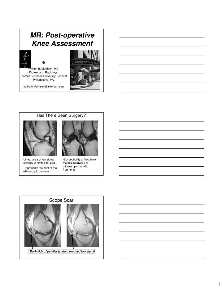
Hyaline Cartilage Surgery
Current Therapies
Cartilage Repair
– OATS – Microfracture – Cartilage transplantation – Proteoglycans???
Kornaat PR, Reeder SB, Koo S, Brittain JH, Yu H, Andriacchi TP, Gold GE. MR imaging of articular cartilage at 1.5T and 3.0T: comparison of SPGR and SSFP sequences. Osteoarthritis Cartilage. 2005 Apr;13(4):338-44. McWalter EJ, Wirth W, Siebert M, von Eisenhart-Rothe RM, Hudelmaier M, Wilson DR, Eckstein F. Use of novel interactive input devices for segmentation of articular cartilage from magnetic resonance images.Osteoarthritis Cartilage. 2005 Jan;13(1):48-53. Roemer FW, Guermazi A, Lynch JA, Peterfy CG, Nevitt MC, Webb N, Li J, Mohr A, Genant HK, Felson DT. Short tau inversion recovery and proton density-weighted fat suppressed sequences for the evaluation of osteoarthritis of the knee with a 1.0 T dedicated extremity MRI: development of a time-efficient sequence protocol.Eur Radiol. 2005 May;15(5):978-87. Epub 2005 Jan 5. Gougoutas AJ, Wheaton AJ, Borthakur A, Shapiro EM, Kneeland JB, Udupa JK, Reddy R. Cartilage volume quantification via Live Wire
- segmentation. Acad Radiol. 2004 Dec;11(12):1389-95.
Watrin-Pinzano A, Ruaud JP, Olivier P, Grossin L, Gonord P, Blum A, Netter P, Guillot G, Gillet P, Loeuille D. Effect of proteoglycan depletion on T2 mapping in rat patellar cartilage. Radiology. 2005 Jan;234(1):162-70. Mosher TJ, Smith HE, Collins C, Liu Y, Hancy J, Dardzinski BJ, Smith MB. Change in knee cartilage T2 at MR imaging after running: a feasibility
- study. Radiology. 2005 Jan;234(1):245-9.
Kornaat PR, Doornbos J, van der Molen AJ, Kloppenburg M, Nelissen RG, Hogendoorn PC, Bloem JL. Magnetic resonance imaging of knee cartilage using a water selective balanced steady-state free precession sequence.J Magn Reson Imaging. 2004 Nov;20(5):850-6.
Or ‘masking’ symptoms
– NSAIDS – Synvisc
MSK radiology research around the world has centered on cartilage imaging
Result: increased emphasis on identification, categorization of cartilage lesions
Autologous Osteochondral Transplantation (AOT)
Autologous bone with overlying cartilage cored from non- weightbearing areas of the joint and transferred into the
Generally indicated for chondral lesions 1.5 – 3 cm
cartilage defect
MosaicPlasty OATS (Osteochondral Autograft Transplant Surgery) SDS (Soft Delivery System) COR Disadvantages- donor site morbidity, limited supply of grafts, long rehab