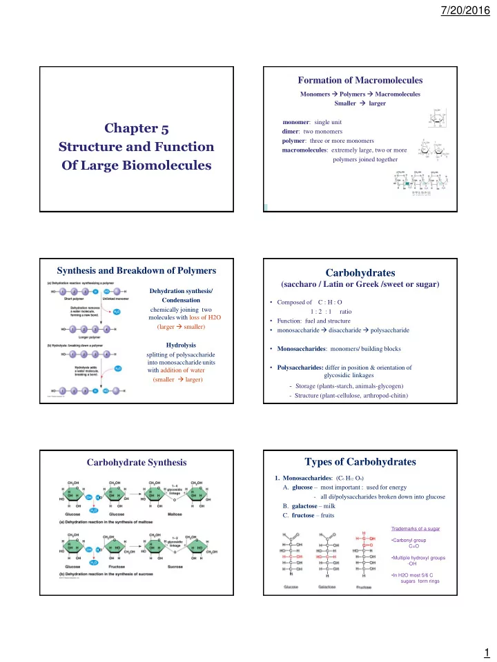

7/20/2016 Formation of Macromolecules Monomers Polymers Macromolecules Smaller larger monomer : single unit Chapter 5 dimer : two monomers polymer : three or more monomers Structure and Function macromolecules : extremely large, two or more polymers joined together Of Large Biomolecules Synthesis and Breakdown of Polymers Carbohydrates (saccharo / Latin or Greek /sweet or sugar) Dehydration synthesis/ Condensation • Composed of C : H : O chemically joining two 1 : 2 : 1 ratio molecules with loss of H2O • Function: fuel and structure (larger smaller) • monosaccharide disaccharide polysaccharide Hydrolysis • Monosaccharides : monomers/ building blocks splitting of polysaccharide into monosaccharide units • Polysaccharides: differ in position & orientation of with addition of water glycosidic linkages (smaller larger) - Storage (plants-starch, animals-glycogen) - Structure (plant-cellulose, arthropod-chitin) Types of Carbohydrates Carbohydrate Synthesis 1. Monosaccharides : (C 6 H 12 O 6 ) A. glucose – most important : used for energy - all di/polysaccharides broken down into glucose B. galactose – milk C. fructose – fruits Trademarks of a sugar • Carbonyl group C=O • Multiple hydroxyl groups -OH • In H2O most 5/6 C sugars form rings 1
7/20/2016 3. Polysaccharides : macromolecules 2. Disaccharides: (C 12 H 22 O 11 ) two monosaccharide units joined (100’s – 1000’s of monosaccharides) by a glycosidic linkage (covalent bond tetween two Cellulose monosaccharides) A. starch – energy storage for plants sucrose – table sugar maltose – malt sugar (beer) - 100’s of glucose molecules B. glycogen – energy storage for animals (muscles and liver) C. cellulose – structure for plant stems - wood and bark (glucose + glucose) - cell walls of plants lactose – milk sugar **Molecules of starch, cellulose, and glycogen- 1000s of glucose units, no fixed size** * function determined by sugar monomers and position of glycosidic linkages* Storage polysaccharides of plants (starch) and animals (glycogen) Cellulose vs. Starch Two Forms of Glucose : glucose & glucose Starch: glucose Cellulose: glucose different geometries = different shapes Lipids (fats) Chitin: structural polysaccharide found in exoskeleton of • not polymers (too small) arthropods. • waxy or oily compounds - differs from cellulose with addition of N • hydrophobic due to H-C chains • ratio of H to C is > 2:1 • structure: 1 glycerol + 3 fatty acids + 3 H2O lost (triglycerol/ide) • functions: - energy storage - membrane formation (phospholipids) - chemical messengers alcohol 16-18 C atoms (sterols/steroids) C- part of carboxyl group (functional group) acidic 2
7/20/2016 Formation of a Triglyceride via Types of Lipids Dehydration Synthesis • Saturated - solid at RT - max number of H bonds with C (saturated with bonds) • Unsaturated - liquid at RT - double bonds between C • Polyunsaturated - many double bonds between C - cooking oils ** trans fats : heating unsaturated fats to become saturated - very bad ** hydrogenated oils : adding H to cis double bonds cause kinks in FA unsaturated fats to make solid - very bad Proteins Phospholipids Steroids Lipid bilayer of cell membrane Cholesterol and hormones Skeleton with 4 fused C rings • Proteios ” = first or primary structure of a phospholipid • 50% dry weight of cells cholesterol • Contains: C, H, O, N, S • Most structurally sophisticated molecules known Functions of Proteins Functions of Proteins 3
7/20/2016 Structure Non-polar Amino Acids • Amino acids - building blocks Amino acid • nonpolar and hydrophobic - 20 different amino acids polymer = polypeptide • protein can be one or more polypeptide chains folded & bonded together • R group (side chain) • large & complex molecules -Variable group -Confers unique chemical complex 3-D shape properties of a.a. • Alpha C is asymmetric Peptide Bond Polar Amino Acids - Covalent bond between two amino acids • polar or charged and hydrophilic - Dehydration synthesis reaction - H from amino group bonds with OH (hydroxyl) of carboxyl group of another amino acid - water molecule is removed - repeated sequence (N-C-C) is the polypeptide backbone Basic Principles of Protein Folding Levels of Protein Structure and Function A. Hydrophobic AA buried in interior of protein • Function depends on structure (hydrophobic interactions) - 3-D structure - twisted, folded, coiled into unique shapes B. Hydrophilic AA exposed on surface of protein (hydrogen bonds) C. Acidic + Basic AA form salt bridges (ionic bonds). D. Cysteines can form disulfide bonds. pepsin collagen hemoglobin 4
7/20/2016 Example of 1 ° Structure Change Primary (1 ° ) Structure Linear chain of amino acids Sickle Cell Anemia Order of amino acids in chain • amino acid sequence determined by gene (DNA) • slight change in amino acid sequence can affect protein’s structure & it’s function • even just one amino acid change can make all the difference! Secondary (2 ° ) Structure Tertiary (3 ° ) Structure • Gains 3D shape (folds, coils) by H bonding • Whole molecule folding - folding along short sections of polypeptide • Bonding between side chains (R groups) of amino acids - Interaction between adjacent amino acids • Hydrophobic interaction: hydrophobic AA in clusters in core Alpha (α) of protein, out of contact with water - helix • van der Waals - H bonding keratin forces hold them between every together 4 th amino acid Beta ( β ) • Anchored by - pleated sheet disulfide bridges - H bonds between (H & ionic bonds) parts of 2 parallel • Include 1 ° and 2 ° polypeptide backbones • structures Core of many globular and fibrous proteins (silk) Quarternary (4 ° ) Structure Models of Tertiary Proteins More than one polypeptide chain joined together. 5
7/20/2016 Review Chaperonins (chaperone proteins) assist in proper folding of proteins • Primary - keep new polypeptide segregated from - aa sequence (peptide bonds) cytoplasmic environment - determined by DNA • Secondary - R groups (H bonds) • Tertiary - R groups ( hydrophobic interactions, disulfide bridges) • Quarternary - multiple polypeptides - hydrophobic interactions Prions : misfolded proteins- mad cow disease Denatration Nucleic Acids - composed of C, O, H, N plus P Unfolding of a protein (disruption of 3 ° structure) - pH, salt, temperature - very large molecules - disrupts H bonds, ionic bonds & disulfide bridges - destroys functionality (sometimes permanently) - polymers of nucleotides (polynucleotides ) DNA RNA ATP 6
Recommend
More recommend