Mitochondrial-Produced Reactive Oxygen Species Matthew Zimmerman, - - PDF document

mitochondrial produced reactive oxygen species
SMART_READER_LITE
LIVE PREVIEW

Mitochondrial-Produced Reactive Oxygen Species Matthew Zimmerman, - - PDF document

6/4/2013 Mitochondrial-Produced Reactive Oxygen Species Matthew Zimmerman, PhD Associate Professor Cellular & Integrative Physiology University of Nebraska Medical Center mczimmerman@unmc.edu Summer 2013 BIOC 998 590 UNL Lecture


slide-1
SLIDE 1

6/4/2013 1

Mitochondrial-Produced Reactive Oxygen Species

Matthew Zimmerman, PhD Associate Professor Cellular & Integrative Physiology University of Nebraska Medical Center mczimmerman@unmc.edu

Summer 2013 BIOC 998‐590 UNL
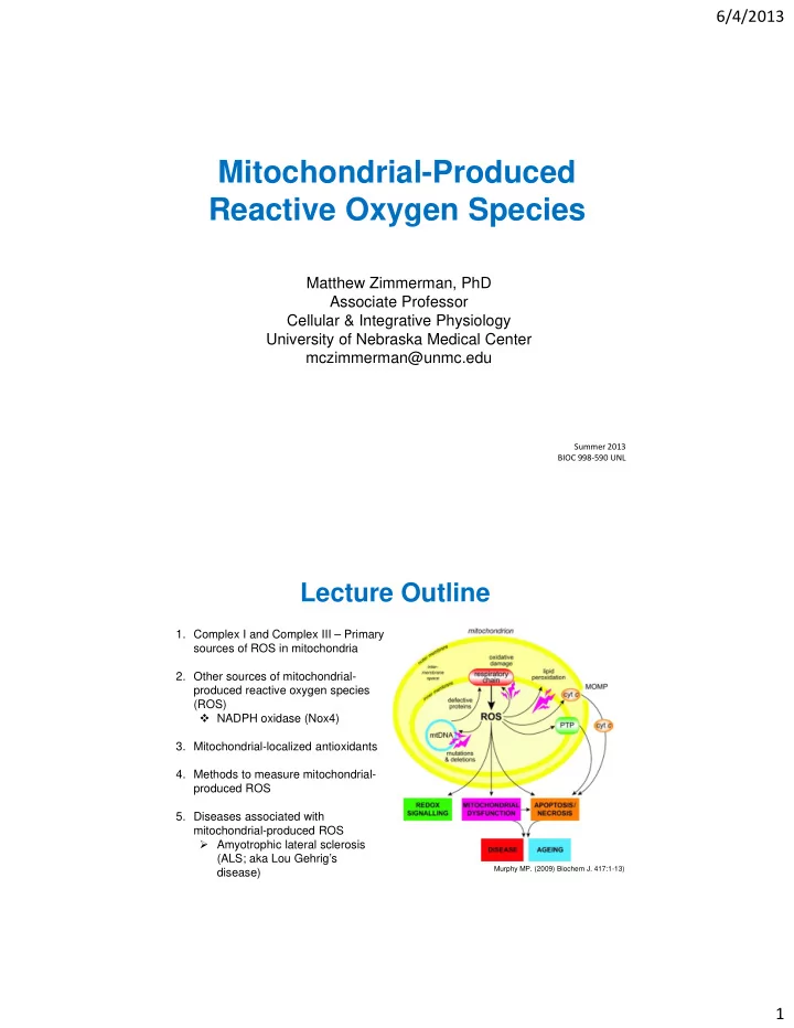
Lecture Outline

  • 1. Complex I and Complex III – Primary

sources of ROS in mitochondria

  • 2. Other sources of mitochondrial-

produced reactive oxygen species (ROS)  NADPH oxidase (Nox4)

  • 3. Mitochondrial-localized antioxidants
  • 4. Methods to measure mitochondrial-

produced ROS

  • 5. Diseases associated with

mitochondrial-produced ROS

  • Amyotrophic lateral sclerosis

(ALS; aka Lou Gehrig’s disease)

Murphy MP. (2009) Biochem J. 417:1-13)

slide-2
SLIDE 2

6/4/2013 2

Sources of Reactive Oxygen Species

  • Mitochondria
  • NADPH oxidase
  • Xanthine oxidase
  • Lipoxygenase
  • Nitric oxide synthases

Turrens JF. (2003) J Physiol. 552.2:335-344

NADPH oxidase

Mitochondrial‐Produced ROS

  • Generally accepted that mitochondrial

energy metabolism is the most quantitatively important source of ROS is most cells

  • Superoxide (O2

‐) is the primary (and

proximal) ROS generated by mitochondria

  • ≈ 0.2 ‐ 2 % of oxygen consumed by

mitochondria is converted to superoxide

  • As electrons flow down chain they can

“leak” off chain on to oxygen → superoxide

  • Presence of SOD in both matrix and

intermembrane space indicates importance

  • f removing O2

‐ from mitochondria

  • MnSOD knock‐out mice are perinatal lethal

Zhang DX. (2006). Am J Physiol. 292:H2023-31)

slide-3
SLIDE 3

6/4/2013 3

Mitochondrial‐Produced Superoxide

  • One-electron reduction of oxygen is thermodynamically favorable for many mitochondrial
  • xidoreductases due to the moderate redox potential of the superoxide/dioxygen couple

Under what conditions can mitochondria electron transport chain (ETC) produced superoxide?

  • 1. Mitochondria not making ATP

and electron carriers are fully reduced

  • 2. High NADH/NAD+ ratio in

mitochondria matrix

  • Can be caused by damage to ETC,

slow respiration, or ischemia

Complex I: A Primary Source of Mitochondrial- Produced Superoxide

Complex I (aka NADH-ubiquinone oxidoreductase; NADH dehydrogenase)

  • Major entry point for electrons into the

electron transport chain (ETC)

  • Flavin mononucleotide (FMN) accepts

electrons from NADH

  • FMN passes electrons to chain of FeS

centers (n=7) and finally to CoQ

  • Produces O2

- from the reaction of oxygen

with the fully reduced FMN (dependent on NADH/NAD+ ratio)

  • Electrons may also leak off FeS centers
  • Inhibition of respiratory chain or increased

levels of NADH increases NADH/NAD+ ratio and, in turn produces O2

-

O2

-

slide-4
SLIDE 4

6/4/2013 4 Complex I: A Primary Source of Mitochondrial- Produced Reactive Oxygen Species

Reverse Electron Transfer (RET) production of superoxide

  • Electrons are transferred against redox potential gradient (reduced CoQ

NAD+)

  • Occurs during low ATP production resulting in a high protonmotive force (p) and

reduced CoQ (succinate or -glycerophosphate supply electrons to reduce CoQ)

  • Rate of RET-dependent superoxide production may be the highest that can occur in

mitochondria

RET: high p and high CoQH2/CoQ

Modified from Murphy MP. (2009)

Complex I: A Primary Source of Mitochondrial- Produced Reactive Oxygen Species

Increasing Complex I-produced superoxide experimentally: Rotenone- induced inhibition of Complex I

  • Rotenone binds to the CoQ-binding site
  • Electrons in Complex I “leak” from either

FMN or FeS centers to oxygen producing superoxide

Modified from Liu Y. et al. (2002). J Neurochem. 780-7.

slide-5
SLIDE 5

6/4/2013 5 Complex III: A Primary Source of Mitochondrial- Produced Reactive Oxygen Species

  • Oxidizes CoQ using cytochrome c as

electron acceptor

  • Reduced CoQ (QH2) transfers one

electron to FeS protein (ISP, aka Rieske protein) and eventually cytochrome c

  • The resulting semiquinone (Q-) transfers

electrons to cytochrome b, then to the Qi site which results in the reduction of another CoQ molecule (Q-cycle)

  • The semiquinone (Q-) is unstable and can

donate electron to oxygen forming superoxide

  • In matrix and intermembrane space

Complex III (aka ubiquinone:cytochrome c reductase)

Modified from Turrens JF, 2003

O2

‐

O2

‐

Complex III: A Primary Source of Mitochondrial- Produced Reactive Oxygen Species

Increasing Complex III-produced superoxide experimentally: Antimycin- induced inhibition of Complex III

  • Antimycin blocks the transfer of electrons to the Qi-site, which results in the

accumulation of the unstable semiquinone

  • The unstable semiquinone can transfer electrons to oxygen producing superoxide

Andreyev A.U., et al. (2005). Biochemistry (Moscow). 70:200-14.

slide-6
SLIDE 6

6/4/2013 6

Additional Sources of Mitochondrial- Produced Reactive Oxygen Species

  • 1. Cytochrome b5 reductase:
  • Outer mitochondrial membrane localization
  • Oxidizes cytoplasmic NAD(P)H
  • Reduces cytochrome b5 in outer membrane
  • May produce O2

- (~ 300 nmol/min/mg protein)

  • Upregulated in schizophrenic patients
  • 2. Monoamine oxidase (MAO):
  • Outer mitochondrial membrane localization
  • Critical in turnover of monoamine

neurotransmitters

  • Catalyze the oxidative deamination of biogenic

amines aldehyde and release of H2O2

  • May be involved in ischemia, aging, Parkinson’s disease

Bortolato M et al. Adv Drug Deliv Rev. 2008

  • 3. Dihyroorotate dehydrogenase (DHOH):
  • Located at the outer surface of inner membrane
  • In the process of pyrimidine nucleotide synthesis, DHOH converts

dihyroorotate to orotate

  • Electron receptor is coenzyme Q and in absence of coenzyme Q produces

H2O2 (in vitro)

  • Role in producing ROS in vivo remains unclear and controversial
  • 4. Dehydrogenase of -glycerophosphate:
  • Located at the outer surface of inner membrane
  • Uses coenzyme Q as electron receptor and catalyzes oxidation of

glycerol-3-phosphate to dihydroxyacetone

  • Studies in mice and drosophila suggest it produces H2O2
  • 5. Aconitase:
  • Localized in matrix
  • Catalyzes conversion of citrate to isocitrate (tricarboxylic acid (TCA) cycle)
  • Inactivated by O2

- and, in turn, produces OH most likely via Fe2+ release

Additional Sources of Mitochondrial- Produced Reactive Oxygen Species

slide-7
SLIDE 7

6/4/2013 7

  • 6. -Ketoglutarate dehydrogenase complex:
  • Located on the matrix side of inner membrane
  • Uses NAD+ as electron acceptor and catalyzes oxidation of -ketoglutarate

to succinyl-CoA

  • Similar to other sources, limited supply of electron acceptor promotes

production of ROS

  • 7. Succinate dehydrogenase (SDH; aka Complex II):
  • Located at the inner surface of

inner membrane

  • Flavoprotein that oxidizes

succinate to furmarate using coenzyme Q as electron receptor

  • Isolated SDH can produce ROS

(again in absence of electron receptor)

  • Mutations in SDH subunits results in an increase in mitochondrial-localized

ROS, particularly superoxide

Modified from Turrens JF, 2003

Additional Sources of Mitochondrial- Produced Reactive Oxygen Species Link between NADPH oxidase and mitochondria?

  • Gp91phox (Nox2) is distributed in the cytoplasm of neurons

and is “particularly abundant near mitochondria”.

Modified from Wang G., et al. 2004 J Neurosci. 24(24):5516-24

NADPH oxidase

slide-8
SLIDE 8

6/4/2013 8

NADPH Oxidase (NOX)‐Derived ROS

  • Multi‐subunit, membrane bound complex that passes electrons through the membrane

from NADPH or NADH to oxygen → superoxide

  • 100x more selective for NADPH over NADH
  • Phagocyte NADPH oxidase first example of a system that produces ROS as the primary

function (not as a byproduct)

  • Responsible for phagocyte respiratory burst
  • Respiratory burst absent in Chronic Granulomatous Disease (CGD) – patients lacking

cytochrome b558 (gp91phox or Nox2 + p22phox)

  • NADPH oxidase subunits include:
  • 7 Nox isoforms (Nox1, Nox2, Nox3, Nox4, Nox5, Duox1, Duox2)
  • Often referred to as the catalytic subunits
  • 2 organizer subunits (p47phox, NOXO1)
  • 2 activator subunits (p67phox, NOXA1)
  • 2 Duox‐specific maturation subunits (DUOXA1, DUOXA2)
  • 1 stabilizing subunit (p22phox)
  • p40phox
  • Active complexes made of a mixture of these subunits

NADPH Oxidase

Conserved Structural Properties Nox Enzymes

  • 1. NADPH binding site in COOH terminus
  • 2. Flavin adenine dinucleotide (FAD) binding

region in COOH terminus

  • 3. Six conserved transmembrane domains
  • 4. Four conserved heme‐binding histidines
  • Electrons are passed from NADPH → FAD →

1st heme → 2nd heme → Oxygen = superoxide

Bedard and Krause, Physiol Rev 2007

O2

‐

slide-9
SLIDE 9

6/4/2013 9

Nox4, NADPH oxidase catalytic subunit, in mitochondria

PNAS, 2009 106(34):14385-14390

Nox4 in neuron mitochondria

Case AJ, et al. Mitochondrial-localized NADPH Oxidase 4 is a Source of Superoxide in Angiotensin II-stimulated Neurons. Am J Physiol Heart Circ Physiol. 2013

slide-10
SLIDE 10

6/4/2013 10

MitoProt Score*: Nox4 – 0.977; MnSOD – 0.985; Prx3 – 0.993; LDH – 0.023; Actin – 0.016

* M.G. Claros, P. Vincens. Computational method to predict mitochondrially imported proteins and their targeting sequences.1996.

  • Eur. J. Biochem. 241, 770-786.

Nox4 in neuron mitochondria

Case AJ, et al. Mitochondrial-localized NADPH Oxidase 4 is a Source of Superoxide in Angiotensin II-stimulated Neurons. Am J Physiol Heart Circ Physiol. 2013

Silencing Nox4 with siRNA attenuates AngII- induced increase in superoxide levels

Case AJ, et al. Mitochondrial-localized NADPH Oxidase 4 is a Source of Superoxide in Angiotensin II-stimulated Neurons. Am J Physiol Heart Circ Physiol. 2013

slide-11
SLIDE 11

6/4/2013 11

Mitochondrial-localized antioxidants

  • 1. Manganese superoxide dismutase (MnSOD, SOD2):
  • Catalyzes dismutation of superoxide producing hydrogen peroxide and oxygen
  • Located exclusively in matrix of mitochondria
  • Nuclear-encoded protein with a mitochondrial-target sequence
  • Homozygous knockout mice only live for few days
  • Large percentage of tumor cells have low MnSOD activity

O2

- + O2 - + 2H+ H2O2 + O2

MnSOD

  • 2. Copper/Zinc superoxide dismutase (CuZnSOD, SOD1)
  • Catalyzes same reaction as MnSOD
  • Primarily found in cytoplasm, but also

present in mitochondria

  • Precise mitochondrial localization is unclear –

most evidence indicates intermembrane space

  • Mechanism of transport into mitochondria

is also unclear

  • Mutant SOD1, associated with

amyotrophic lateral sclerosis (ALS), appears to accumulate in mitochondria

Zhang DX. (2006). Am J Physiol. 292:H2023-31)

CuZnSOD is commonly thought of as the cytoplasmic localized SOD, but it is also expressed in mitochondria

slide-12
SLIDE 12

6/4/2013 12

Mitochondrial CuZnSOD in neurons following adenovirus-mediated gene transfer

Control (Non-infected) AdCuZnSOD (50MOI, 96h) MitoTracker Red CuZnSOD Merged

Mitochondrial CuZnSOD protein and activity are increased following gene transfer

EM micrograph of Mito-fraction

1 2 3 1 2 3

  • 1. Control (Non-infected)
  • 2. AdEmpty
  • 3. AdCuZnSOD

Whole cell lysate Mito-fraction

MnSOD LDH (Cytosolic marker) COXIV CuZnSOD MnSOD CuZnSOD (human) CuZnSOD Calnexin (ER marker)

(Mito markers)

slide-13
SLIDE 13

6/4/2013 13

Mitochondrial-localized antioxidants

  • 3. Glutathione
  • ~ 10% glutathione levels in cells is in mitochondria
  • Can be transported into mitochondria via specialized GSH-transporters
  • Oxidized glutathione (GSSG) can be reduced back to GSH by glutathione

reductase localized in the matrix

  • 4. Glutathione peroxidase (GPx1)
  • Uses GSH for the reduction of hydrogen peroxide to water
  • Found in mitochondrial matrix and intermembrane space
  • 5. Phospholipid glutathione peroxidase (PhGPx; GPx4)
  • Reduces lipid hydroperoxides and hydrogen peroxide
  • GPx4 long form expressed in mitochondria
  • Knockout mice are embryonic lethal
  • 6. Cytochrome C
  • Present in intermembrane space
  • Can scavenge superoxide
  • The reduced cytochrome c is recycled by cytochrome c oxidase
  • Biological significance of cytochrome c as a superoxide scavenger in vivo

remains to be fully elucidated

Mitochondrial-localized antioxidants

  • 6. Peroxiredoxins (Prx)
  • Reduce hydrogen peroxide and

lipid hydroperoxides

  • Prx3 highly expressed in heart,

adrenal gland, liver and brain mitochondria

  • Prx5 highly expressed testis
  • 7. Thioredoxin (Trx) system
  • Trx2 recycles Prx by reducing the

disulfide

  • Oxidized Trx2 is then recycled by

thioredoxin reductase (TrxR), which uses NADPH as the source

  • f reducing equivalents

Echtay KS. (2007) Free Rad Biol Med. 43:1351-71

slide-14
SLIDE 14

6/4/2013 14

Smith R.A.J., et al. (2008) Ann NY Acad Sci. 1147:105-111

Exogenous mitochondrial-targeted antioxidants

  • Antioxidant compounds covalently attached to a lipophilic

triphenylphosphonium cation target mitochondria

  • Such compounds include: SOD mimetic M40403 (MitoSOD)

and tempol (MitoTempol); peroxidase mimetic ebselen (MitoPrx); coenzyme Q (MitoQ); tocopherol (MitoE)

Methods for measuring mitochondrial-produced ROS

  • 1. MitoSOX Red fluorescence
  • Mitochondrial-targeted superoxide

sensitive fluorogenic probe (Invitrogen/Molecular Probes)

  • MitoSOX is dihydroethidum (DHE; aka

hydroethidine) linked to a triphenylphosphonium group

  • Like DHE, fluorescence can be

detected using 405, 488, 510 nm excitation

  • However, only the 405 nm

excitation detects the 2-hydroxyethidium fluorescent product which is specifically dependent on superoxide

Dikalov S. et al. (2007). Hypertension

slide-15
SLIDE 15

6/4/2013 15

Methods for measuring mitochondrial-produced ROS

  • 2. Electron paramagnetic resonance (EPR) on isolated mitochondria
  • Mitochondria can be isolated from tissue or cultured cells
  • Isolated mitochondria incubated with EPR spin trap or spin probe
  • Amount of spin trap/probe radical are detected using EPR

Important issues to consider:

  • Purity and integrity of mitochondria preparation
  • Depending on spin trap/probe selected use specific

antioxidants (e.g. SOD) to selectively measure a particular ROS

Mariappan N. et al. Free Rad Biol Med. (2009). 46:462-70.

Methods for measuring mitochondrial-produced ROS

  • 3. Amplex Red to detect hydrogen peroxide efflux from isolated mitochondria
  • Isolated mitochondria incubated with Amplex Red in the presence of HRP
  • Measure levels of fluorescent product, resorufin

Important issues to consider:

  • Purity and integrity of mitochondria preparation
  • If using this method to indirectly measure superoxide, remember not all

superoxide is converted to hydrogen peroxide (nitric oxide in mitochondria)

  • Numerous matrix peroxidases will consume hydrogen peroxide
slide-16
SLIDE 16

6/4/2013 16

Summary

1. Numerous sources of ROS production in mitochondria 2. Complex I and III have been the most studied and best characterized, and, to date, are generally considered the primary sources of mitochondrial-produced ROS 3. Nox4 has also been reported to be a source of ROS in mitochondria 4. Collection of mitochondrial-localized antioxidants also play a significant role in the levels of ROS in mitochondria 5. Mitochondrial superoxide can be elevated experimentally with rotenone (Complex I inhibitor) or antimycin A (Complex III) inhibitor 6. Mitochondrial ROS can be reduced experimentally by using antioxidant compounds linked to a triphenylphosphonium group or by increasing expression of endogenous antioxidant proteins

  • Fatal neurodegenerative disease that specifically

targets motor neurons in the spinal cord, brain stem, and cortex

  • Most common adult motor neuron disease; 5,600

cases diagnosed each year in U.S.

  • Disease onset usually begins with weakness in arms

and legs and quickly progresses to total paralysis

  • Patients generally die of respiratory failure 2‐5 years

after the first symptoms appear

  • ALS often referred to as Lou Gehrig’s disease

Amyotrophic Lateral Sclerosis (ALS)

From alsa.org

slide-17
SLIDE 17

6/4/2013 17

  • 20-25% of familial ALS cases are associated

with mutations in a cellular antioxidant enzyme called CuZnSOD (SOD1)

  • Mutant CuZnSOD-induced neuronal toxicity is

believed to involve a toxic gain of function; not a loss of SOD activity

  • Many familial ALS mutant CuZnSOD

proteins retain SOD activity

  • CuZnSOD knockout mice do not develop

motor neuron disease

  • Overexpressing wild-type CuZnSOD in

animal or cell culture models of ALS does not provide protection

  • Mutant CuZnSOD expression is ubiquitous,

although only motor neurons appear to be affected

From Valentine JS, 2005.

  • Annu. Rev. Biochem
  • 1. Control
  • 2. Wild‐type CuZnSOD
  • 3. Mutant CuZnSOD #1
  • 4. Mutant CuZnSOD #2
  • 5. Mutant CuZnSOD #3
  • 6. Mutant CuZnSOD #4

Amyotrophic Lateral Sclerosis (ALS) Expression of different CuZnSOD mutants in cultured neurons decreases cell survival

slide-18
SLIDE 18

6/4/2013 18

Mutant CuZnSOD increases superoxide levels in mitochondria Overexpressing MnSOD attenuates mutant CuZnSOD-mediated increase in mitochondrial superoxide

slide-19
SLIDE 19

6/4/2013 19

Overexpression of MnSOD (SOD2) inhibits mutant CuZnSOD-mediated neuronal toxicity Intramuscular injection of AdSOD2 results in retrograde transport and SOD2 expression in spinal cord motor neurons

Control AdSOD2

slide-20
SLIDE 20

6/4/2013 20

Intramuscular injection of AdSOD2 delays motor dysfunction in ALS transgenic mice