
SLIDE 1
Infection Prevention Boot Camp I for the Novice Infection Preventionist January 16‐17, 2020 Florida Hospital Association | Mission to Care Hospital Improvement Innovation Network 1
Microbiology
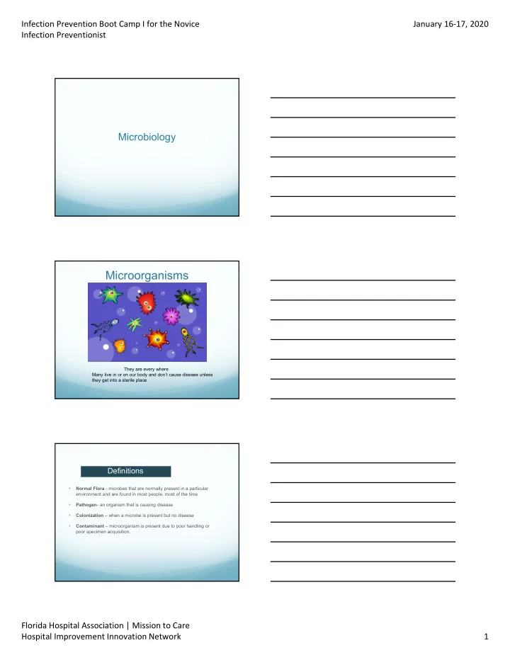
Microorganisms
They are every where Many live in or on our body and don’t cause disease unless they get into a sterile place
Definitions
- Normal Flora - microbes that are normally present in a particular
environment and are found in most people, most of the time
- Pathogen- an organism that is causing disease
- Colonization – when a microbe is present but no disease
- Contaminant – microorganism is present due to poor handling or
poor specimen acquisition.