Medical and Family History Neurological Exam Past Medical and - - PowerPoint PPT Presentation

medical and family history neurological exam
SMART_READER_LITE
LIVE PREVIEW

Medical and Family History Neurological Exam Past Medical and - - PowerPoint PPT Presentation

2/10/2017 CC: Difficulties with gait and slurred speech 59 y.o. RH Caucasian female August 2015 : Back pain and left leg pain -Progressive loss of balance and frequent falls Recent Advances in Neurology -Changes in speech and handwriting


slide-1
SLIDE 1

2/10/2017 1

Liliana Ramirez Gomez M.D. Assistant Clinical Professor of Neurology February 10, 2017

Recent Advances in Neurology Difficult Diagnosis

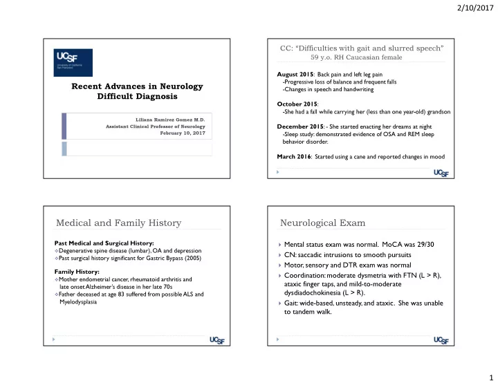
CC: “Difficulties with gait and slurred speech”

59 y.o. RH Caucasian female August 2015: Back pain and left leg pain

  • Progressive loss of balance and frequent falls
  • Changes in speech and handwriting

October 2015:

  • She had a fall while carrying her (less than one year-old) grandson

December 2015: - She started enacting her dreams at night

  • Sleep study: demonstrated evidence of OSA and REM sleep

behavior disorder. March 2016: Started using a cane and reported changes in mood Past Medical and Surgical History:

Degenerative spine disease (lumbar), OA and depression Past surgical history significant for Gastric Bypass (2005)

Family History:

Mother endometrial cancer, rheumatoid arthritis and

late onset Alzheimer’s disease in her late 70s

Father deceased at age 83 suffered from possible ALS and

Myelodysplasia

Medical and Family History

Mental status exam was normal. MoCA was 29/30 CN: saccadic intrusions to smooth pursuits Motor, sensory and DTR exam was normal Coordination: moderate dysmetria with FTN (L > R),

ataxic finger taps, and mild-to-moderate dysdiadochokinesia (L > R).

Gait: wide-based, unsteady, and ataxic. She was unable

to tandem walk.

Neurological Exam

slide-2
SLIDE 2

2/10/2017 2

August 2015: Normal CBC, CMP , TSH, B12 and vitamin D level October 2015: EMG/NCS lower limbs were normal December 2015: Sleep study Moderate degree of sleep apnea and REM sleep behavior disorder January 2016: EEG normal awake

Previous work up (outside studies)

Brain MRI August 2015 C spine MRI August 2015 Differential diagnosis

Acquired causes Genetic causes Idiopathic causes

Neurodegeneration Dominant Recessive Mitochondrial Metabolic X linked Other Nutritional Endocrine Metabolic Infectious Inflammatory Neoplastic Paraneoplastic Autoimmune Toxic Modified from Fogel et al 2011.

slide-3
SLIDE 3

2/10/2017 3

Based on the history what do you think is the most likely etiology in this case?

A.

Metabolic/nutritional/toxic

B.

Neurodegenerative

C.

Genetic

D.

Immune mediated: autoimmune or paraneoplastic

E.

Demyelinating disease

M e t a b

  • l

i c / n u t r i t i

  • n

a l / t

  • x

i c N e u r

  • d

e g e n e r a t i v e G e n e t i c I m m u n e m e d i a t e d : a u t . . . D e m y e l i n a t i n g d i s e a s e

23% 27% 14% 33% 4%

Tempo of Illness as a Clue to Diagnosis

Fogel and Perlman, 2011 Fogel et al. JAMA Neurol. 2014;71(10):1237-1246

Brain MRI March 2016

slide-4
SLIDE 4

2/10/2017 4

Brain MRI March 2016

August 2015 March 2016 August 2015 March 2016

C spine MRI March 2016

slide-5
SLIDE 5

2/10/2017 5 Cerebellar peduncles hyperintensities

Vascular: vascular malformations or stroke Infectious: PML, Lyme disease, Listeria rhombencephalitis,

Whipple

T

  • xic/Traumatic: Heroin induced leukoencephalopathy

Autoimmune: neurosarcoid, vasculitis (e.g., Bechet's), CLIPPERS Metabolic: osmotic demyelination syndrome Inherited/Genetic: Fragile X associated tremor-ataxia syndrome Neurodegenerative: MSA-C Neoplastic: lymphoma or glial neoplasms Demyelinating: MS or ADEM

Fragile X-associated tremor/ataxia syndrome Multiple system atrophy of cerebellar type (MSA-C)

slide-6
SLIDE 6

2/10/2017 6

Based on the radiological findings what do you think is the most likely diagnosis in this case?

A.

MSA C

B.

Primary progressive MS

C.

Fragile X-associated tremor/ataxia syndrome

D.

An autoimmune or paraneoplastic disorder

MSA C Primary progressive MS Fragile X-associated tre... An autoimmune or para...

33% 31% 27% 10%

REM Sleep Behavior Disorder and RBD associated disorders

α-synucleinopathies Narcolepsy Secondary to medications (SSRIs, SNRIs) Others:

Wilson’s disease, PSP, FTD, ALS, MS, brainstem infarction, subarachnoid hemorrhage VGKC antibody-associated limbic encephalitis.

Courtesy of Dr. Lisa Ashbrook

Laboratory work up

Hematologic and metabolic work up was normal

Vitamin E, Thiamine, Serum copper Heavy metals in urine (lead, mercury, arsenic): negative VLCFA, plasma amino acids, urine organic acids were nl Pyruvate: 0.98 nl AFP: 3.9 nl

Genetic Fragile X testing showed 30/22 CGG repeats Infectious: RPR, HIV, Lyme, Hep B and C negative Autoimmune: ESR, RF, ANA, SSA, SSB, cANCA, pANCA TTG, anti gliadin Ab were all negative

slide-7
SLIDE 7

2/10/2017 7

CSF 3/24/16 and 6/13/16

RBC 3 WBC 2 Glucose 52/63 Protein 38/37 IgG index: normal 2 unique oligoclonal bands in CSF were detected compared to serum

  • --Repeat LP in June showed no OGB

ACE level in CSF 0.9 nl (0-2.5) Lyme IgG, IgM: no bands detected VDRL negative Serum paraneoplastic panel: (Ataxia panel at Mayo x2) Patient tested positive for P/Q-type calcium channel (VGCC) antibodies 0.15

Fogel and Perlman, 2011

P/Q-type voltage-gated calcium channel (VGCC)

Nachbauer et al. J Neurol (2015) 262:1385–1393

PETCT CTAC WHOLE BODY

slide-8
SLIDE 8

2/10/2017 8 Treatment

Outcome measure: Scale for the assessment and rating of ataxia (SARA)

Course of IV pulse

steroids x 5 days

High dose prednisone for

2 months with a slow taper

Course of IVIG

Responses to and Outcomes of Treatment

  • f Autoimmune Cerebellar Ataxia in Adults

118 patients from the Mayo clinic with autoimmune ataxia Median age at onset was 58 years, 73.7% were women 63 patients had paraneoplastic and 55 patients had

nonparaneoplastic ataxic disorders.

Improvements were significantly more common among

patients with nonparaneoplastic disorders and those with exclusively PMP antibodies

Jones AL , McKeon et al. JAMA Neurol. 2015;72(11):1304-1312

What would you do next?

A.

Try a different immunosuppressive treatment

B.

Genetic testing

C.

Advise a “watch and wait” approach

D.

Counsel that nothing else can be done

Try a different immunos... Genetic testing Advise a “watch and wai.. Counsel that nothing els...

72% 2% 17% 10%

Additional immunosuppressive treatment

IV pulse cyclophosphamide 15 mg/kg per CYCLOPS protocol (used in vasculitis)

Harper L, et al. Ann Rheum Dis. 2012 Jun;71(6):955-60

slide-9
SLIDE 9

2/10/2017 9 Genetic testing

Complete evaluation for autosomal dominant and

recessive panel associated with ataxia

Dominant: none Recessive:

A pathogenic heterozygous frameshift mutation for SYNE1 A heterozygous missense mutation as a variant of unknown

significance on SYNE1 (unclear if on same allele)

SYNE1 mutations in autosomal recessive cerebellar ataxia

Mutations in the synaptic nuclear envelope protein 1

(SYNE1) gene, located on chromosome 6p25, were first reported in patients from a province of Quebec, Canada.

Autosomal recessive cerebellar ataxia type I, is a slowly

progressive ataxia that leads to moderate disability and diffuse cerebellar atrophy on brain imaging.

With a relative frequency of ∼5%, SYNE1 is one of the

more common recessive ataxias worldwide

Synofzik et al. Brain. 2016 May;139(Pt 5):1378-93

Refined differential diagnosis and treatment

Differential:

Autoimmune cerebellar Ataxia: positive VGCC +P/Q Neurodegenerative MSA C SYNE1 mutation may play a role

Treatment:

Immunosuppressive therapy Periodic surveillance for malignancy Supportive care

Acknowledgments

Thank you to Dr. Jeff Gelfand at UCSF We welcome your referrals at the:

General Neurology Clinic 400 Parnassus Ave., Eighth Floor San Francisco, CA 94143 Phone: (415) 353-2273 Fax: (415) 353-2898