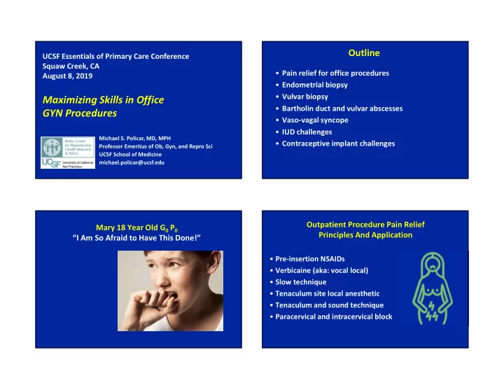
Maximizing Skills in Office Bartholin duct and vulvar abscesses GYN - - PowerPoint PPT Presentation

maximizing skills in office
SMART_READER_LITE
LIVE PREVIEW

Maximizing Skills in Office Bartholin duct and vulvar abscesses GYN - - PowerPoint PPT Presentation

Outline UCSF Essentials of Primary Care Conference Squaw Creek, CA Pain relief for office procedures August 8, 2019 Endometrial biopsy Vulvar biopsy Maximizing Skills in Office Bartholin duct and vulvar abscesses GYN Procedures


slide-1
SLIDE 1

Maximizing Skills in Office GYN Procedures

Michael S. Policar, MD, MPH Professor Emeritus of Ob, Gyn, and Repro Sci UCSF School of Medicine michael.policar@ucsf.edu

UCSF Essentials of Primary Care Conference Squaw Creek, CA August 8, 2019

Outline

  • Pain relief for office procedures
  • Endometrial biopsy
  • Vulvar biopsy
  • Bartholin duct and vulvar abscesses
  • Vaso-vagal syncope
  • IUD challenges
  • Contraceptive implant challenges

Mary 18 Year Old G0 P0 “I Am So Afraid to Have This Done!”

  • Pre-insertion NSAIDs
  • Verbicaine (aka: vocal local)
  • Slow technique
  • Tenaculum site local anesthetic
  • Tenaculum and sound technique
  • Paracervical and intracervical block

Outpatient Procedure Pain Relief Principles And Application

slide-2
SLIDE 2

Non-Steroidal Anti-inflammatory Drugs

Cochrane review, 2015

  • Tramadol and naproxen had some effect on

reducing IUD placement pain in specific groups

  • Lidocaine 2% gel, misoprostol, and most NSAIDs did

not help reduce pain

  • Conventional wisdom

–Rx naproxen sodium 550 mg or Ibuprofen 800 mg –Helps mainly with post-placement cramping

Lopez LM et al. Interventions for pain with IUD insertion. Cochrane Database of Systematic Reviews 2015, Issue 7. Art. No.: CD007373

Verbicaine

  • Keep her talking!
  • Calm, soothing vocal tone
  • Slow, easy pace
  • Utilize whatever works for the patient ASK
  • Breathing techniques
  • Mindful mediation
  • Guided imagery

Distraction Language Considerations

Blending compassion, medical fact, and positive suggestion

“Most patients are worried about pain, and they are often surprised when it is easier than they had

  • expected. As we proceed, let us

know how you are feeling so that we can make adjustments. We want this to go well for you.”

slide-3
SLIDE 3

Language Considerations…

“Relax” You might feel “a pinch”

  • r ”a stick and a burn”

“You’re doing great”

“Try taking a deep breath”

“You might feel a sensation

  • r “a twinge”

“You might feel a sensation

  • r “a twinge”

“It’s a natural reaction to lift up. See

the table.”

“It’s a natural reaction to lift up. See

if you can let your hips be heavy on the table.” “I can see you’ve had practice with relaxation”

Try: Instead of:

Language Considerations…

  • I’m going to put a

grasper on your cervix

  • Now I’m going to

sound your uterus

  • Here comes the

inserter “You may notice three cramps, then we’ll be done”

Try: Instead of:

Let me know if you want me to tell you before each one

Tenaculum Choose Site for Placement

  • Anterior lip
  • Posterior lip
  • Typically a horizontal bite, some prefer vertical

Tenaculum: Size of Bite

  • 1-1.5 cm wide
  • 1 cm deep
  • Not too shallow- may tear through
  • Not too deep- unnecessary
slide-4
SLIDE 4

Tenaculum Pain Reduction

  • Once the teeth are in contact with the cervix,

press into the tissue

  • Close the tenaculum very, very slowly

– Only to the first or second stop – Silently

  • Once the ratchet is closed, test your application

gently to be sure it is secure

Tenaculum Pain Reduction

  • Some providers recommend injection of 1cc local

anesthetic at the tenaculum site – Have patient cough or use other distraction

  • Don’t move the tenaculum inadvertently
  • Hook fingers thru rings to place tenaculum
  • During sounding and IUD placement, don’t hook

your fingers through the rings…hold the shank

Palm Up Middle finger in ring Palm Up Ring Finger in Ring

slide-5
SLIDE 5

1-2 Clicks Still with Palm Up – Ring Finger Ring Finger Still Palm Up So You Can See Above Your Hand!

Tenaculum Use When Sounding

  • Change hands; hold the tenaculum with the non-

dominant hand while sounding and for placement

  • OK to let tenaculum lay on speculum when picking

up the sound or IUD

  • Thumb on one side of ratchet; fingers on the other

– Avoid the rings – Avoid inadvertent movements

Fingers NOT in rings

slide-6
SLIDE 6

Can “Choke up” Uterine Sound: Purpose

  • Ensure that you can pass through the internal os
  • Direction and pathway through the os to the fundus
  • Measures depth/distance from external os to fundus

–Appropriate for IUD placement not <5.5 cm –10 cm or more in some cases –Tells you where to set the flange –So you don’t waste the IUD

Like a Dart

slide-7
SLIDE 7

Or Like Pencil Uterine Sound Pain Reduction

  • If metal; bend sound to mimic uterine flexion
  • Hold it like a pencil or dart
  • Use wrist action

–Not elbow –Not shoulder

  • Brace fingertips on speculum to achieve control of

force while advancing the sound

Uterine Sound Pain Reduction

S-l-o-w Progression

  • Through the internal os
  • Pause once when through the internal os
  • Slow intentional progression to the fundus
  • Avoid momentum

Uterine Sound Pain Reduction

  • Touch the fundus once

–Repeated tapping is unnecessarily uncomfortable for the patient

  • Move slowly and intentionally

–Moving too quickly increases discomfort

  • If difficulty sounding, consider

–EMB sampler

slide-8
SLIDE 8
  • Place paracervical or intracervical block at any point
  • Use a thinner sound (endometrial sampler)
  • Use os finder device
  • Dilate internal os with small metal or plastic dilator
  • Try a shorter wider speculum
  • Reposition the tenaculum onto a different place
  • If unsuccessful, return after misoprostol 200 mg per

vagina 10 hours and 4 hours prior to placement

Still Unable To Pass Through Internal Os Os Finder Device

Cervical Os Finders (Disposable Box/25) Cervical Os Finder Set (Reusable Set of 3)

Dilators

  • Dilate internal os with metal dilators
  • #13 french

– Divide by 3.16 to get mm (4.1 mm)

  • Double ended
  • Tapered ends ease passage through os
  • Choke up on the handle
  • Sterile lubricant on tip
  • Leave the (small) sound in the canal and come

alongside the sound with the inserter

Passed Through with Sound …But not the inserter!

slide-9
SLIDE 9

Cervical Anesthesia

10-20 ml of 1% lidocaine (NO epinephrine)

Carrie Cwiak, MD, MPH 10 20 30 40 50 60 70

Median Pain Scores w/ 20 mL 1% Lidocaine

No PCB PCB

Pain with IUD Placement Lidocaine Block vs. Sham Block

Mody et al. ObGyn 2018

Pain with IUD Insertion, Nulliparas

Akers et al. ObGyn 2017

10 mL

Paracervical Block

  • Target is uterosacral ligaments, which contain the

cervical and uterine nerves

  • Use spinal needle OR 25g, 1 ½” needle + extender
  • Inject at reflection of cervico-vaginal epithelium
slide-10
SLIDE 10
  • 5-10 cc 1% lidocaine (no epinephrine) each side
  • Submucosal injection 5mm-1cm deep
  • Short speculum allows more movement
  • WAIT 1-2 minutes after placing block

Paracervical Block Paracervical Block Paracervical Block

X X X 4 o’clock 8 o’clock

slide-11
SLIDE 11

X X 4 o’clock 8 o’clock 6 o’clock X X X

Paracervical Block Intracervical Block

  • Targets the paracervical nerve plexus
  • 1 ½ inch 25g needle with 12 cc

“finger lock” syringe

  • Inject ½- 1 cc. at 12 o’clock, then apply tenaculum

Intracervical Block

  • Angulate needle at the hub to 45o lateral direction
  • At 3 o’clock, insert needle into cervix to the hub 1

cm lateral to external os, then aspirate –Inject 4 cc of local, then 1 cc while withdrawing

  • Rotate barrel 180o, then inject at 9 o’clock
slide-12
SLIDE 12

Intracervical Block

X 8 o’clock X 6 o’clock X 9 o’clock 3 o’clock 4 o’clock

Lidocaine Safety

  • Inject in correct spot
  • Aspirate to avoid intravascular injection
  • Metallic taste is a common side effect

Maximum Local Anesthetic Dosing

Local Anesthetic

Onset

(mins)

Max Dose (mg/kg)

without/with epi

Max Dose (mg)

without/with epi

55kg pt dose

without/with

Lidocaine 4-7 4.5/7 mg/kg 300/500 mg 25/38 mL Bupivacaine 10-20 2.5 mg/kg 175 mg 55 mL Chloroprocaine fast 11/14 mg/kg 800/1000 mg 60/77 mL

  • Rough estimates that are not evidence-based
  • Lower peak levels and slower absorption with vasoconstrictor
  • Adding bicarb (to lidocaine) speeds onset of action
  • Bupivacaine with less difference since med is vasoconstrictive

Endometrial Biopsy

slide-13
SLIDE 13

Who Needs an EMB?

  • Purpose: detect endometrial hyperplasia or cancer
  • Menopausal woman

– Any postmenopausal bleeding, if not using HT – Unscheduled bleeding on continuous-sequential hormone therapy – Bleeding > 3 mo after start of continuous-combined hormone therapy – Endometrial stripe > 5 mm (applies to postmenopausal woman only) – Pap smear: any endometrial cells or AGC Pap

Who Needs an EMB?

Premenopausal Women

  • Prolonged metrorrhagia
  • Unexplained post-coital or intermenstrual bleeding
  • Endometrial cells on cytology in an anovulatory

premenopausal woman

  • Atypical Glandular Cells (AGC) cervical cytology

– Abnormal endometrial cells, or – Older than 35 years old – Under 35 yo with abnormal bleeding

Technique of EMB

  • Bimanual exam to evaluate uterine axis, size
  • Cleanse cervix with antiseptic
  • S-l-o-w-l-y apply tenaculum ( + local anesthetic)
  • Use of the sampling device

– Choose correct type (rigidity) of sampler – “Crack” stylet to ensure easy movement – Gently advance to fundus; expect resistance at internal os – Note depth of sounding with side markings – Pull back stylet to establish vacuum

slide-14
SLIDE 14

Technique of EMB

  • Use of the sampling device (continued)

— Rotate in a helical direction from the fundus to the os in

  • rder to use the lateral cutting edge of the port

— If the sampler has filled, remove  place tissue in fixative — If the sampler did not fill, repeat 2-3 more passes — If a “curette check” for completeness is desired, perform in- and-out motion in vertical strips to confirm a “gritty” feel — Cut tip of sampler and empty any remaining tissue

  • Remove the tenaculum; check for bleeding
  • Remove the speculum
  • Move the patient to a supine position for a few minutes

Tips for Internal Os Stenosis

  • Pain relief

– Use para-cervical or intra-cervical block – Intrauterine instillation of lidocaine

  • Cervical dilation

– Freeze endometrial sampler to increase rigidity – Grasp sampler with ring forceps 3-4 cm from tip – Use cervical “os finder” device – Use small size Pratt or Hegar dilators – No evidence to support misoprostol priming

Indications for Vulvar Biopsy

  • Papular or exophtic lesions, except obvious condylomata
  • Thickened lesions (biopsy thickest region) to differentiate

VIN vs. LSC

  • Hyperpigmented lesions (biopsy darkest area), unless
  • bvious nevus or lentigo
  • Ulcerative lesions (biopsy at edge), unless obvious herpes,

syphilis or chancroid

  • Lesions that do not respond or worsen during treatment
  • In summary: biopsy whenever diagnosis is uncertain

Vulvar Biopsy

slide-15
SLIDE 15

Tools for Vulvar Biopsy

Photo courtesy of Dr Hope Heafner

  • Insulin syringe
  • 1% lidocaine with or

without epinephrine

  • 2x2 or 4x4 gauze sponge
  • Unsterile exam gloves
  • Antiseptic solution (e.g,

povidone-iodine or chlorhexidine

  • Silver nitrate sticks or

Monsel’s solution

  • Pathology container and

label

Tips for Vulvar Biopsies

  • Where to biopsy

– Homogeneous : one biopsy in center of lesion – Heterogeneous: biopsy each different lesions

  • Prep skin with antiseptic
  • Skin local anesthesia

– Most lesions will require ½ cc. lidocaine or less – Epinephrine will delay onset, but longer duration – Use smallest, sharpest needle: insulin syringe – Inject anesthetic s-l-o-w-l-y

  • Alternative: 4% liposomal lidocaine (30 minutes) or EMLA

(60 minutes) pre-op

  • Stretch skin; twist 3 or 4 mm Keyes punch back-and-forth

until it “gives” into fat layer

Tips for Vulvar Biopsies

  • Lift circle with forceps or needle; snip base
  • Hemostasis with AgNO3 stick or Monsel’s solution
  • Silver nitrate will not cause

a tattoo

  • Suturing the vulva is almost

never necessary

  • Separate pathology container

for each area biopsied

  • LABEL the container!!!
slide-16
SLIDE 16

Bartholin Duct and Vulvar Abscess Management

from: Omole F, Am Fam Physician 2003

Bartholin’s Duct and Gland Conditions

  • 2% lifetime risk of developing BD cyst or abscess, especially

during reproductive years

  • BD abscess is 3-times more common than BD cyst
  • If duct becomes blocked or transected

– No infection: BD cyst – Primary infection: acute BD cellulitis or abscess – Rarely, BD cyst is secondarily infected  abscess – All surgical treatments are designed to drain fluid and create a new duct

  • BG/BD carcinoma is rare; occurs in women > 40 yo

Bartholin Duct Cellulitis

(aka: Bartholinitis, Bartholin adenitis)

  • Painful red induration of lateral perineum at 5 or 7 o’clock,

but no palpable abscess

  • Most commonly due to skin streptococcus
  • Treatment

– Cephalexin 500 mg PO QID or – Clindamycin 300-450 mg PO QID – 5 day course, but extend if not improved (IDSA #15) – Moist heat: sitz baths, warm compresses

slide-17
SLIDE 17

Bartholin Duct Cellulitis

  • Re-evaluate in 2-4 days

– Cellulitis will either have improved or point as abscess – If abscess develops, perform I&D

  • Admit immunocompromised women (especially diabetics)

for IV antibiotics and close observation – Risk of developing necrotizing fasciitis

Bartholin Duct Abscess

  • Develops over 2-4 days; up to 8 cm diameter
  • Tend to rupture and drain after 4-5 days
  • Pain may range from local discomfort to severe pain

– BD abscess can be so painful that the patient is incapacitated; difficulty in walking or sitting

  • Physical exam

– Fever present in one-third of patients – Acutely tender swelling at posterior labium majora extending inwards into the base of labium minora – Occasionally track anteriorly up L majora (Rouzier, 2005) BD Abscess Pre-treatment

Pregnant, diabetic, or immunocompromised? Yes Admit No Large enough to drain? No Yes Moist heat Abx if induration RTC in 48-72 hours Resolved Abscess points Tolerates manipulation No Conscious sedation in ED or office Yes I&D Word catheter

Pus: C/S + GC/Ct if STD risks

BD Abscess: I&D Tools and Supplies

  • Povidone-iodine solution
  • Anesthetic solution (1-2% lidocaine) + insulin needle/syringe
  • Word catheter (diameter: #10F Foley catheter)

– 22-25 gauge needle and 5 mL-syringe, plus water or gel, for inflation of catheter tip

  • No. 11 blade scalpel
  • Hemostat (for breaking up loculations)
  • Saline solution for irrigation
  • Collection kits for bacterial culture and GC/Ct NAAT
slide-18
SLIDE 18

BD Abscess: Tips for Word Catheter

  • Consider topical skin anesthetic with EMLA
  • Have assistant retract abscess laterally to select incision

site…immediately external to the hymeneal ring

  • Inject skin with 1-2 cc. lidocaine
  • 5-10 mm. stab with # 11 blade perpendicular to abscess
  • Gently lyse loculations with clamp
  • Irrigate cavity with saline
  • Insert needle into Word port; then test the bulb
  • Insert Word catheter; inflate (3-5cc) until snug fit in cavity
  • Tuck nipple into vagina

Word Catheter: Correct Position BD Abscess: Post-Drainage Management

  • Sitz baths and warm compresses for 2-3 days
  • Antibiotics not needed routinely after I&D
  • If residual cellulitis, SIRS, or immunocompromise,

recommended antibiotic regimens include Strep: Cephalosporin (cephalexin or cefixime) And for Staph: TMP/SMX 1-2 double strength tablets PO BID

  • r Doxycycline 100 mg PO BID

– If MRSA confirmed, replace doxycycline with TMP/SMX

slide-19
SLIDE 19

Treatment of Vulvar Abscess

  • Abscess <2 cm with mild cellulitis

– Moist heat: sitz-bath, warm compresses – 1st line: TMP-SMX 1-2 DS tabs BID for 5-10 days – 2nd line: doxycycline (100 mg BID) or

  • Clindamycin (300-450 mg TID)

– Follow-up one week later

  • Abscess >2 cm or less than 2 cm and present > 1 week

– I&D, with packing if possible – Aerobic c/s for MRSA – Follow-up at 2 days and 2 weeks after treatment

Incision & Drainage

  • Dome infiltration with

local anesthetic

  • in A-P axis, incise point

with #11 blade

  • Send culture
  • Break up loculations
  • Irrigate
  • Pack as needed
  • Saline-soaked gauze replaced daily until the defect has closed

Chen K, UpToDate. 2016

  • Extensive or rapidly progressing surrounding cellulitis
  • Abscess size ≥5 cm
  • Location makes abscess difficult to drain completely
  • Infection extends into other anatomic compartments (e.g.,

abdominal wall or thigh)

  • High likelihood of MRSA
  • Systemic signs of infection
  • Immunocompromised patient
  • Recurrent abscess

Vulvar Abscess: I&D, then Antibiotics Treatment of Vulvar Abscess With Cellulitis

  • I&D, then antibiotics and serial surveillance
  • Antibiotics

– Staph: TMP/SMX or doxycycline PLUS – Strept: cephalosporin or clindamycin

  • 5-10 days of therapy is recommended

– Duration of therapy guided by resolution of symptoms

slide-20
SLIDE 20

Betsy 17 year old G0

  • While having her LNg IUD placed, Betsy

says, “Is this going to take much longer? I really need to go to the bathroom”

  • What’s going on here??

Betsy 17 year old G0

  • She recalls after the fact that she had a

fainting spell after her HPV immunization

  • She had told her PCP about this

problem…heart auscultation and an ECG were normal.

Vasovagal Response, Episode Or Attack AKA: Non-cardiogenic Syncope

  • Mechanism

– Starts with peripheral vasodilation – Bradycardia + drop in B/P

  • More likely with
  • Pain with cervical manipulation
  • Previous episodes of vaso-vagal fainting
  • Dehydration or NPO

Grubb BP N Engl J Med 2005

Presyncopal Signs

  • Facial pallor (distinct green hue)
  • Yawning
  • Pupillary dilatation
  • Nervousness
  • Diaphoresis
  • Slurred or confused speech

Grubb BP N Engl J Med 2005

slide-21
SLIDE 21

Presyncopal Symptoms

  • Weakness/light-headedness
  • Visual blurring/tunnel vision
  • Nausea
  • Feeling warm or cold
  • Sudden need to go to the bathroom
  • Tinnitus

Grubb BP N Engl J Med 2005

Vasovagal Prevention

  • Good hydration (electrolyte/ sports drink)
  • Eat before placement
  • Prophylactically contract muscles if known

history

Grubb BP N Engl J Med 2005

How to Abort a Vasovagal

  • Isometric contractions of the extremities
  • Intense gripping of the arm, hand, leg and

foot muscles

  • No need to bring the legs together or change

position– just tense the muscles

  • These contractions push blood back into the

center of the body

  • ….and abort the reflex

Missing strings

slide-22
SLIDE 22

Missing String…Possibilities

  • 1. IUD in-situ

– String coiled in canal or endometrial cavity – String short, broken, or severed

  • 2. Unnoticed expulsion
  • 3. Intrauterine pregnancy

Malpositioned IUD, following perforation or incorrect placement

  • 4. Embedment into the myometrium
  • 5. Translocation into the abdomen or pelvis

Missing String…Possibilities

Do not use the white stabilizing rod as a plunger during placement of a copper IUD

Prevention of Perforation: Copper IUD

Missing String: Office Ultrasound

  • No IUD string in canal
  • Pregnancy test negative
  • Office ultrasound (UTZ)

Desires removal Extract + guidance KUB Leave In Situ Absent Hysteroscopy Present Expelled Absent Present 3D-UTZ or CT with contrast Present Translocated Extracted Embedded Desires retention Not found Absent “Formal” UTZ Embedded? Laparoscopy 3D-UTZ or CT with contrast

  • r
slide-23
SLIDE 23

Missing String: No Office Ultrasound

  • No IUD string in canal
  • Pregnancy test negative

Extracted Attempt extraction Desires removal Embedded Not felt Desires retention Ultrasound KUB In Situ Absent KUB Op hysteroscopy Present Translocated Absent Expelled Extracted Absent Present Ultrasound Absent Translocated In Situ OR

Missing String: Desires Removal

Extraction of IUD in-situ

  • 1. Consent for uterine instrumentation procedure
  • 2. Bimanual exam
  • 3. Probe for strings in cervical canal
  • 4. Apply tenaculum
  • 5. Administer cervical block
  • 6. Choose extraction device

– Emmett Thread Retriever – Patterson alligator forceps – Ring IUD: crochet hook or 3-5 mm suction curette

Emmett Thread Retriever Thread Retriever

slide-24
SLIDE 24

Fulcrum 1 cm from the tip of the device Opened and closed completely within the uterine cavity No cervical dilation necessary

Prabhakaran S, Chuang A, Contraception 2011.

Missing String: Desires Removal

Extraction of IUD in-situ

  • 7. Intrauterine exploration for a T-shaped IUD

– Real-time ultrasound guidance may help, if available – Gently open/ close/quarter turn forceps at progressive depths until “purchase” of stem or arm

  • 8. Maneuver hook along anterior, then posterior, uterine wall

from fundus to canal

  • 9. If embedment suspected, consider evaluation with 3-D

ultrasound or pelvic CT with contrast – Extract via operative hysteroscopy or laparoscopy

Why Do CT or 3-D Ultrasound?

Answer: To decide whether to start the extraction with laparoscopy or hysteroscopy!

Missing String: Desires Removal

Additional measures, as indicated

  • Pain management

– Cervical block + oral NSAIDs for pain – Conscious sedation

  • Cervical dilation

– Osmotic dilator – Rigid dilators – Misoprostol may facilitate IUD extraction

slide-25
SLIDE 25

Identify the Insertion Site

  • Inner side of non-dominant upper arm
  • Overlying the triceps muscle about 8-10

cm (3-4 inches) from the medial epicondyle of the humerus

  • 3-5 cm (1.25-2 inches) posterior to the

sulcus (groove) between the biceps and triceps muscles

Implant Location After Insertion

Old site New site

  • This location is intended

to avoid the large blood vessels and nerves lying within and surrounding the sulcus.

  • If it is not possible to

insert the implant in this location (e.g., in women with thin arms), it should be inserted as far posterior from the sulcus as possible.

Implant Insertion Troubleshooting

  • If you pierce the skin, retract and re-insert subdermally
  • If the implant protrudes from the insertion site, remove it

and perform a new procedure with a new implant

  • If the rod is not palpable…

– Check the applicator (purple tip of the obturator should be visible) – Use imaging (x-ray, CT, ultrasound, MRI) – Until location is confirmed, counsel to use other method

  • Deep implants need to be removed to prevent migration

Implant Removal Tips

  • Only attempt removal if you have localized it

– Identify radiologist who can identify it on u/s – Obtain u/s in your clinic – Can also obtain etonogestrel level if not radio-opaque

  • If you can feel it, you can often remove it

– Fine mosquito clamps are key

  • Identify referral center for deep removals

– It takes special expertise if below the muscle fascia