Learning points Superficial lower limb venous anatomy Clinical - - PDF document

learning points
SMART_READER_LITE
LIVE PREVIEW

Learning points Superficial lower limb venous anatomy Clinical - - PDF document

19/08/2019 Learning points Superficial lower limb venous anatomy Clinical assessment in OPD Varicose veins Endovenous Assessing suitability for EVT therapy Understanding the procedure US imaging after EVT Prof Tim


slide-1
SLIDE 1

19/08/2019 1

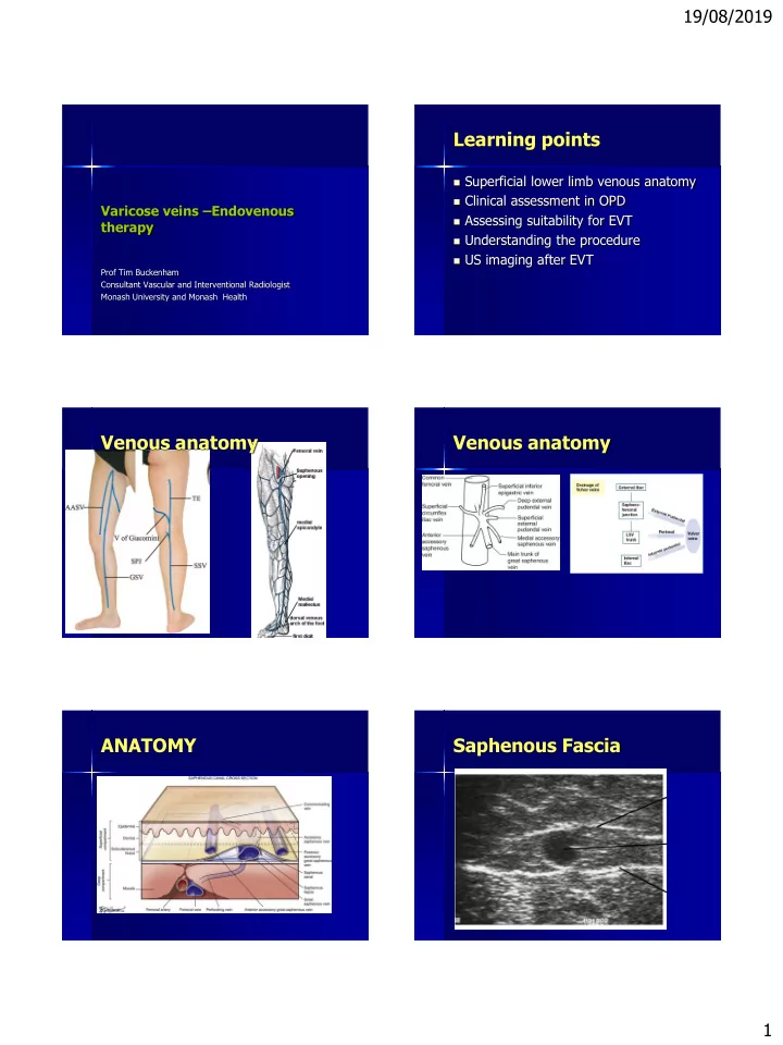
Varicose veins –Endovenous therapy

Prof Tim Buckenham Consultant Vascular and Interventional Radiologist Monash University and Monash Health

Learning points

 Superficial lower limb venous anatomy  Clinical assessment in OPD  Assessing suitability for EVT  Understanding the procedure  US imaging after EVT

Venous anatomy Venous anatomy ANATOMY Saphenous Fascia

slide-2
SLIDE 2

19/08/2019 2

Varicose veins the physiology Perforating veins

Relationship between Vulval varicosities Lower limb varicosities and Pelvic congestion

OVARIAN VEIN INCOMPETENCE

  • During pregnancy, the vascular capacity of the
  • varian veins may increase 60-fold and remain

this way for months after delivery.

  • Left ovarian vein usually affected

Trans abdominal US

 Dilated Ovarian

vein(>6 mm)

 Reverse flow  Incompetent  Para ovarian varices

Treatment of Incompetent ovarian vein

slide-3
SLIDE 3

19/08/2019 3

The Team

Vascular surgeons, Nurses, Sonographers, IR’s, PSA

Physical requirements:

 Good quality diagnostic ultrasound facilities with

tilting table

 Trained sonographers  Experienced IR nurses  Consultation rooms

EVT techniques

  • 1. EVLT
  • 2. RF ablation
  • 3. Cyanoacrylate closure
  • 4. UGS

Interventional room:

 Ultrasound machine  Tilting table  Sterile facilities  Good lighting

CEAP classification of chronic venous disease

Clinical classification

C0 No visible or palpable signs of venous disease

C1 Telangiectasies or reticular veins

C2 Varicose veins

C3 Edema

C4a Pigmentation or eczema

C4b Lipodermatosclerosis or athrophie blanche

C5 Healed venous ulcer

C6 Active venous ulcer

  

complications

slide-4
SLIDE 4

19/08/2019 4

Advantages of EVT

 Ambulatory procedure  No GA  No incision  Immediate activity/work after

procedure

 At least as effective as surgery

Patient Flow

 Procedure time laser 30-40min/limb  UGS 15/min limb  Application of full length class 2

compression hosiary

 Ambulatory recovery period 30

minutes

Patterns of disease and their treatment

Great and Small saphenous incompetence:

Can be treated with EVT Contraindications to EVT:

1.

Non-occlusive thrombus

2.

Tortuosity

3.

Dilatation > 10mm(relative)

4.

Deep Venous incompetence/obstruction

GSV Imaging prior to EVT

 GSV patent  GSV in the saphenous sheath for at

least 15cm

 Is the GSV cirsoid?  Does GSV have webs  Diameter of GSV

Neo Junction

 Possible to treat

with EVLT

 If there is a trunk

slide-5
SLIDE 5

19/08/2019 5

ENDOVENOUS

 Percutaneous access seldinger  Sheath advanced over a wire to 5mm

short of junction (US control) plus light

 Tumescent local  Withdraw laser/RF probe

Tip of laser within vein GSV tumescent local EVLT V RFA

slide-6
SLIDE 6

19/08/2019 6

Laser safety Managing the AASV Sclerotherapy

 1ml of 3% Fibrovein foamed to 4mls  Tessario technique  Maximum dose 8mls of foam  US guided

3% fibro vein and Tessario foam

Complications

 Scotoma  Pigmentation  Ulceration  Eccyhmosis

3 days continuous and 3 weeks during daytime

slide-7
SLIDE 7

19/08/2019 7

Conclusion

IR can set up a venous service Utilise existing infrastructure provided by an existing Radiological department with skilled IR nurses Attractive option for patients and Hospital

1.

Outpatient treatment

2.

Minimally invasive

3.

No GA

4.

No surgery

5.

Very rapid recovery