
SLIDE 1
in Prior Asbestos Workers using Low-Dose Computed Tomography - - PowerPoint PPT Presentation
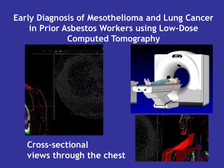
Early Diagnosis of Mesothelioma and Lung Cancer in Prior Asbestos Workers using Low-Dose Computed Tomography Cross-sectional views through the chest nodule pleural plaques pleural plaques: reliable marker for asbestos exposure
pleural plaques:
thickness area / volume density calcifications? involved pleura (costal, diaphragmatic, mediastinal) symmetry? fluid? shape (flat or lobulated)
Baseline low-dose CT
no or inconspicuous plaques
no or non-specific nodules annual repeat no change bi-annual repeat indeterminate nodules
(≥5 mm solid or ≥8 mm non-solid)
suspicious plaques
lobulated, asymmetric, effusion
6 months f/u no change annual repeat suspicious nodules (≥15 mm)
with effusion immediate biopsy
endobronchial nodules 3 months f/u resolved (mucous) annual repeat stable bronchoscopy growth biopsy etc.
Lung Car 20% Pleural 13% Peritoneal 6% Other malignancy 61% Distribution of 64 Malignancy in 1287 participates Colon cancer Prostate cancer Kidney Hemopathy Esophageal cancer Bladder cancer Pancreas cancer Liver cancer Breast Duodenal Throat cancer 6 3 7 3 7 4 3 1 1 1 1
Other Malignant 39