Idiopathic Intracranial Hypertension w ith Papilla Edem a & - - PowerPoint PPT Presentation

idiopathic intracranial hypertension w ith papilla edem a
SMART_READER_LITE
LIVE PREVIEW

Idiopathic Intracranial Hypertension w ith Papilla Edem a & - - PowerPoint PPT Presentation

Is there any correlation betw een Idiopathic Intracranial Hypertension w ith Papilla Edem a & Intracranial Venous Disorders? Judit Som lai* , Bernadett Salom vry ** * Unit of Neuro-Ophthalm ology , Department of Neurology & Stroke


slide-1
SLIDE 1

dr@Som laiJudit.hu www.Som laiJudit.hu/ en Judit Som lai*, Bernadett Salom váry**

*Unit of Neuro-Ophthalm ology,

Department of Neurology & Stroke Military Hospital, Budapest, Hungary * ** Unit of Neuro-Ophthalm ology National Institute of Clinical Neuroscience, Budapest, Hungary

Is there any correlation betw een

Idiopathic Intracranial Hypertension w ith Papilla Edem a & Intracranial Venous Disorders?

slide-2
SLIDE 2
  • 1. Reversed way of thinking is needed in Neuro- Ophthalm ology

because Eye sym ptom s precede and pre-indicate the disorders of Central Nervous System

  • 2. The neuro - ophthalm ological syndrom es help us to find

the exact etiology of the IIH syndrom e w ith „ unknow origin” In case of vision loss caused by IIH There is no m orphological sign of disorders on MRI *

  • 3. In case of a negative MRI: Neuro-Opthalm ological exam inations show

the Topographic Localisation of the optic nerve’s disorders *

  • 4. The earlier w e diagnose the disease, the better chance

to prevent an irreversible visual loss

OCULAR SYMPTOMS – as a precursor

  • f

CSF +/ - VENOUS circulatory disease

slide-3
SLIDE 3

How rapidly will the optic nerve diseases

– with different pathogenesis -

cause significant visual loss ?

slide-4
SLIDE 4

The long and com plex history of diagnostic criteria

  • f IIH classification

Friedm an DI, Jacobson DM (2002). "Dia gnostic criteria for id iop a thic intra cra nia l hy p ertension".

  • Neurology. 59 (10): 1492–1495.(20 0 2)

A CHRONOLOGY

  • f the im portant landm arks

in defining the CRITERIA system of the IIH Modified version of the Dandy PTC criteria (the latest classification

  • f IIH)

Quincke (18 90 )

  • „m eningitis serosa”- headache, visual loss, papilledema
  • etiology: hypersecretion of CSF

LP: liquor pressure > 25 water cm s

& liquor without biochemical and cytological disorders Nonne (190 4)

  • First called as „pseudotum or cerebri” (PTC)

Dandy (1937)

  • Dandy’s criteria of PTC
  • Modification of the Dandy criteria (1982)

Dandy WE (October 1937). Annals of Surgery. 106 (4): 492–513.

Clinical sym ptom s of HIP:

Headeache Vom iting Transient visual obscurations Papilledem a No focal neurological signs

(exception: paresis of the abducens nerve) Foley (1955)

  • „Benign Intracranial Hypertension - BIH „

Corbett et al. (198 2)

  • „Idiopathic Intracranial Hypertension” (IIH)

Sm ith JL (198 5).

  • "Whence pseudotum or cerebri?".

J of Clinical Neuroophthalm ology. 5 (1): 55–6. 198 5.

No typical signs of the Higher Intracranial Pressure (HIP) on CT/ MRI * Norm al CT/ MRI findings without evidence of IC throm bosis Unknown etiology of HIP

slide-5
SLIDE 5

Com parison of the diagnostic criteria

  • f

HIP & IIH in the background of Papilla Edem a

Higher Intracranial Pressure (HIP)

in the b a ckground of p a p illa ed em a

The latest classification of

Intracranial Idiopathic Hypertension

(IIH) (A m odified version of the Dandy PTC criteria) Significant elevation

  • f

Intracranial Pressure >>> 25-30 m m Hg LP: liquor pressure > 25 water cm s Monro-Kellie principles: 1. Incom pressible:

Brain - CSF - Blood (Vbrain+V CSF+V blood=constant)

  • 2. Disproportion inside the rigid cranium ;

No typical signs of the HIP

  • n CT/ MRI

Norm al CT/ MRI findings without evidence of throm bosis CAUSES of the HIP:

  • Intracranial tum our
  • CSF secretion-, CSF circulatory-,

&/ CSF flow disorder

  • Bra in ed em a
  • IC v enous circula tory d isord er

CSF reabsorption problem – sinus thrombosis

Clinical sym ptom s of HIP: Headeache Transient Visual Obscurations Papilledem a Unknown etiology of HIP !

slide-6
SLIDE 6

Higher Intracranial Pressure (HIP) Causes

Relationship betw een :

CSF circulation & IC venous circulation

Matthew J. Thurtell et coll. An Update on Idiopathic Intracranial Hypertension Rev Neurol Dis. 20 10 Spring-Summer; 7(0): e56–e68.

1. Cerebrospinal Fluid (CSF)

secretion circulation hydrocephalus resorption

Higher IC Pressure (HIP)

CSF : reabsorption disorders

Norm al Pressure Hydrocephalus (NPH)

SSS throm bosis stenosis of SSS CSF : flow- circulation diseases

  • bstructiona l hydrocephalus

(IC tum our)

CSF : secretion disorder

hy p ersecretiona l hydrocephalus (m eningitis)

  • 3. IC venous circulatory disorders

CSF reabsorption problem : NPH Sa gitta l Sup erior sinus(SSS)

stenosis/ throm bosis

  • 4. Primary Intracerebral

Venous Circulatory Disorders

  • 2. Disproportion between

the volum e + content of cranium intracranial tum our developm ental disease

  • f HIP?

&

  • f IIH?

What is the actual cause

slide-7
SLIDE 7

Where to find the reabsorption disorders within the SSS?

IIH : etiopathom echanism – theories in the background of Papilla edem a CSF production & circulatory CSF - liquor absorption

CSF pressure= Rout CSFform ation+Psss+Vascular com ponent?

Cerebral Venous Circulatory system

https:/ / courses.lumenlearning.com/ boundless- ap/ chapter/ protection-of-the-brain/

Diseases: HYDROCEPHALUS

  • hypersecretional
  • obstructional

Diseases: liquor absorption disorders Non-resorptive HYDROCEPHALUS CSF ABSORPTION =(PCSF – PSSS) ROUT

Conrad E Johansson et al. Multiplicity of cerebrospinal fluid functions: New challenges in health and disease Cerebrospinal Fluid Res. 20 0 8 ; 5: 10.

Diseases:

  • IC VENOUS

MALFORMATION

  • SINUS THROMBOSIS

CSF pressure - manometer LP - shunt: LP, VP

King, J.et al.. Cerebral venography and m anom etry in IIH. Neurology, Vol. 45, No. 12, ( 1995),

slide-8
SLIDE 8

What leads to Throm bosis in the Cerebral Venous System ?

IIH:etiopathom echanism – theories : in the background of papilledem a

Com pression of venous sinuses

Stenosis and throm bosis

  • f venous sinuses

Slowing and stasis of the IC venous flow

The increase of the IC venous blood pressure

CSF – reabsorption decrease HIP with brain swelling

Venous and arterio-capillar intravascular pressure increases.

Brain infarct

Syndrom e Groups IC Venous Network Circulatory Disorders Throm bosis

  • f Cerebral Sinuses
  • Cavernosus Sinus (SV)
  • Sagittal Superior Sinus (SSS)
  • Transversal Sinus (ST)

Norm al Pressure Hydrocephalus

CSF- reabsorption disorders – caused by

SSS stenosis/ stasis, throm bosis

Throm bosis in Multiple Sinus

slide-9
SLIDE 9

Where to look for the diseases within the Intracerebral Venous Circulatory System

w hen the patient has uni-, or bilateral papilledem a?

CEREBRAL

venous circulatory system

INTRACEREBRAL network

superficial veins-cortex deep veins of substance DURAL SINUS system

Posterior - Superior

SSS; SSI, s. rectus,

  • s. transversus,
  • s. sigm oideus,s. tentorialis,
  • s. occipitalis

Anterior - Superior

sinus cavernous, s.intracavernous

  • s. parietalis, s. basilar,
  • s. sphenoparietal, s. petrosal:sup, inf.

Farb RI et al. IIH: The p rev a lence a nd m orp hology of sinov enous stenosis. Neurology V. 60, No. 9, (20 0 3), p.1418-24.

OCULAR

venous blood supply system

ORBITAL VENOUS SYSTEM

  • v. ophthalmica superior (VOS)
  • v. ophthalmica inferior (VOI)

CENTRAL RETINAL VEIN connection with cavernosus sinus Physiological relationship between CSF circulatory & IC venous circulatory

to cavernous sinus

slide-10
SLIDE 10

10

The Results of an International FERRO Study On Cerebral Vein and Dural Sinus Throm bosis (ISCVT) Prognosis of Cerebral Vein and Dural sinus throm bosis (624 pts.)

José M. Ferro , Patrícia Canhão, Jan Stam, Marie-Germaine BousserB and Fernando Barinagarrementeria;

  • Stroke. 20 0 4 ;35:664-70

headache: 553 (8 8 %) * papillaedem a: 174 (28 ,3%)

* visual loss: 8 2 (13,2%) double vision 8 4 (13,5%) Occluded

sinuses / veins Pts. No.

(of 624)

%

SAGGITAL SUPERIOR sinus 313 62,0 Lateral Sinus left 279 44,7 Lateral Sinus right 257 4 1,2 Straight Sinus 112 18 ,0 Deep Venous System 6 8 10 ,9 Cortical veins 10 7 17,1 Jugular veins 74 11,9 Cerebellar veins 3 0 ,3 CAVERNOUS Sinus 8 1,3

slide-11
SLIDE 11

Clinical Ocular Signs draw our attention to the location of the Cerebral Venous Throm bosis throm bosis

  • f

CEREBRAL VEINS throm bosis

  • f

IC-SINUSES HIGHER INTRACRANIAL PRESSURE

(liquor absorption disorders & venous flow disorders)

OPHTHALMIC vein

pre Retinal trunk throm bosis branch CORTICAL veins VISUAL FIELD defect

CAVERNOUS sinus:

  • chemosis,
  • phthalmoparesis
  • exophthalmos
  • retinal prethrombosis
  • periferal eye-movement disorders

SAGITTAL SUPERIOR Sinus PAPILLEDEMA hemiparesis, epilepsy, coma TRANSVERSAL Sinus (otitis purulent) PAPILLEDEMA paresis n.VI. +retroorbital pain

Thrombosis of Sagittal Superior Sinus & Parietal veins : PAPILLEDEMA

slide-12
SLIDE 12

Throm bosis of

venous sinuses

Underlying illness as Causes

CAVERNOUS SINUS Septic inflam m ation

  • nasal & paranasal sinusitis
  • orbital infection

Aseptic diseases

  • cranial trauma,
  • facial operation,

SAGITTAL SUPERIOR SINUS Dural Arteriovenous fistula

(after recanalisation of sinus thrombosis)

Cranial traum a (vertex ) * Prothrom bosis Tum our

  • parasagittal

meningeoma (recidivous)

  • meningeal cc.

TRANSVERSE & SIGMOID SINUS Mastoiditis

  • straight to the sinus
  • by vv. Emissaria
  • pr. Hem atology

syndrom e &

  • sec. Coagulopathy

Gradenigo

Syndrom e

( purulent otitis, abduction paresis retroorbital pain)

INTERNAL JUGULAR VEIN

  • surgical
  • traumatic

Tum ours

  • intravascular
  • extravascular

Which Background Illnesses can cause certain sinus throm boses?

slide-13
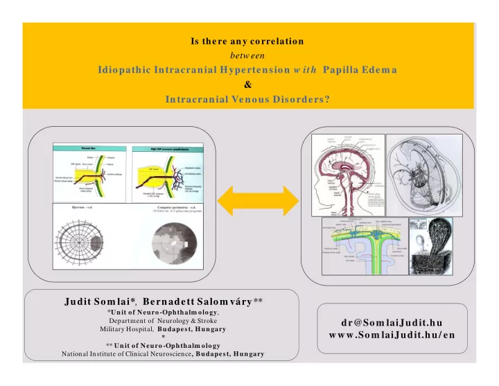
SLIDE 13

System ic & Neurological Illnesses of CNS

Congenital throm bophilia

FV (Leiden) m utation, hyperhom ocysteinaem ia, FIIG20 210 A polym orphism us, AT, PC-, PS deficiency, „sticky platelet syndrome”

Acquired throm bo- phylia

polycythem ia, Antiphospholipid syndrom e, m alignant disorders pregnancy, oral contraceptives Paroxysmal nocturnal haemoglobinuria (PNH), hyperhomocysteinaemia , cryofibrinogenemia, , throm bocytosis gynecological diseases – postpregnancy, colitis, Chron disease nephrotic sy., thyreotoxicosis m ed ica tions: ovarium hyperstimulation syndr., androgens, antioestrogenic

Abnorm alities

  • f

blood flow

com pression: meningeoma, glomus npl., lymphoma, metastasis cathetherization, dehydration, congenitalis heart diseases, persistant pulm onary hypertension, Dural AVM

Abnorm alities

  • f

vessel walls

  • local infections
  • traum a
  • after surgical intervention (embolisation of AVM)
  • carcinom atous infiltration

Collection of System ic & Neurological Illnesses of CNS that leads to Intracerebral Throm bosis

* Walsh-Hoyt: Clinical Neuro-Ophthalm ology , Venous Occlusive Disease. 6th ed.p.2445

slide-14
SLIDE 14

Transient Visual Obscuration: (TVO)

(prognosis of the initial papilledema)

  • uni / bilateral
  • >>>> 1’
  • without visual loss
  • pulsatile tinnitus

Big Blind Spot sign

(the most sensitive predictor of papilledema)

  • Functions of the optic nerve remain intact for some time
  • Later, contraction of the visual field

Chronic Papilledem a

Untreated or undertreated cases OPTIC ATROPHY– chronic congestion PAPILLEDEMA – optic atrophy-decrease of visual acuity

Sym ptom s – Papilledem a The ea rliest & m ost significa nt sign of IIH

Cello KE et al,. Fa ctors a ffecting v isua l field outcom es in the id iop a thic intra cra nia l hy p ertension trea tm ent tria l. J Neuroophthalm ol. 20 16;36:6–12-

slide-15
SLIDE 15

Sym ptom s - Eye Movem ent Disorders

  • papilledema is preceded by double vision -

15

Paresis of abduction nerve - diplopia Horizontal Abduction Paresis

  • in prim ary position: convergent strabism us
  • cover test: esophoria > esotropia
  • horizontal gaze paresis
  • with/ -out vertical skew deviation
  • upward gaze paresis ( Parinaud sy.)

Clinical syndromes: Horizontal gaze paresis ‘One and a half’ Syndrom e Parinaud syndrom e

slide-16
SLIDE 16

Sym ptom s - Neurological signs w ith Papilledem a

16

Headache:

  • bifrontal, in the morning
  • >80-90%

* Neurological focal signs

(at a latest stages of the illness)

  • m onoparesis, hem iparesis
  • epileptic seisure
  • central vom itus

In childhood: acute –fulminant signs :

caused by: m ore exp ressed ed em a d isp osition „sunset sign”– upward ophthalm oplegy w ith ataxia, facial paresis, neck stiffness, torticollis

slide-17
SLIDE 17

Tests of Visual Function History Ophthalm oscopy

test (+ / - )

How to find the Cerebral Venous Throm bosis -

through a sequence of Exam ination ? - algorythm -

Differential Diagnosis of Papilledem a

PAPILLA

norm al/ decoloration atrophy PAPILLEDEMA

yes no

Macula

yes

OPTIC NERVE LESIONS Inflam m ation Ischaem ia Com pression

  • Tumour
  • CSF - HIP

bilateral

  • Papilledem a (HIP)
  • IC Sinus

Throm bosis * unilateral Retinal praethrom bosis

VISUAL LOSS?

Afferent pupillom otoric reflex

STLY=V?

Am sler grid OCT

STLY=V?

  • H. Wilhelm et al. Visual Loss of uncertain origin:

Diagnostic Strtategies. From : U. Schiefer et al. Clinical Neuro-Ophthalm ology,. A practical Guide. 6-7. Springer Verlag, 20 0 7.

slide-18
SLIDE 18

Entity - Syndrom e Sym ptom s - Finding Decision

Testing of the Afferent pupillom otoric reflexes(RAPD) Testing

  • f the Visual Function

History Pathological Ophthalm oscopy

Diagnostic algorithm

for the Evaluation of Visual Disorder caused by Papilla edem a

Functional testing Morphological testing Additional testing

Visual Field test

Pinhole

(defect of fixation?)

L ADDITIONAl exam inations (+/ - etiology : uncertain ) tünet

1. m acular - papillom acular- areas papillar -

  • 2. prechiasm al-

chiasm al- n.II. retrochiasm al-

  • 3. additional m ethods
  • H. Wilhelm et al. Visual Loss of uncertain
  • rigin: Diagnostic Strtategies.

From : U. Schiefer et al. Clinical Neuro-Ophthalm ology, A practical Guide. 6-7. Springer Verlag, 20 0 7.

slide-19
SLIDE 19

DIAGNOSTICS

  • f the Optic Nerve System

FUNCTIONAL

exam ination m ethods of the ON

Basic exam inations:

  • Visual acuity : for D & N
  • Colour vision, Am sler grid - test,
  • Afferent Pupillomotoric pathway - RAPD

Exam inations of the conductivity of Optic Nerves

  • Critical Fusion Frequency (CFF) test
  • Electrophysiology : ERG-, VEP

Visual Field tests:

  • Confrontation VFT
  • Cam pim etry with tagent:

Bjerrum screen

  • Com puter perimetry: OCTOPUS

Blood flow exam inations:

  • Heidelberg Retinal Flowmeter (HRF)
  • Fluorescein angiography (FLAG)

Michael Wall et al., The NORDIC IIH Study Group; Visual Field Outcom es for the Idiopathic Intracranial Hypertension Treatm ent Trial . Invest Ophthalm ol Vis Sci. 20 16 Mar; 57(3):805–12.

Ophthalm oscopy: by direct &/

  • r indirect funduscopy

Morphological m easurem ent of the:

GCC of the Macular- & Papillom acular Regions

  • f the ON

Optical Coherence Tom ography (OCT) analysis of Ganglion Cell Com plex: GCC: RNFL+ GCL+IPL (loss of layers thickness)

Optical Coherence Tom ography Substudy Com m ittee and the NORDIC Idiopathic Intracranial Hypertension Study Group. Papilledem a outcom es from the optical coherence tom ography substudy

  • f the idiopathic intracranial hypertension treatm ent trial.

Ophthalm ology. 20 15;122:1939–1945.

>>>

MORPHOLOGICAL

exam ination m ethods of the ON

Log Reflection Log Reflection Optic Disk Optic Disk Fovea Fovea RNFL RNFL Choroid Choroid Vitreous Vitreous Sclera Sclera 250 µm 250 µm 250 µm
slide-20
SLIDE 20

DIAGNOSIS

  • f Eye Movem ent disorders

Unconscious pts.

  • Primary eye position
  • Pupillomotoric reflex

Conscious patients

  • Primary eye position
  • Pupillomotoric reflex
  • smooth pursuit eye movements (9dir)
  • analysis of the double images

Analysis of Double Vision

  • Near double images test

Maddox wing

  • Distant double images test

Hess screen, Polatest

  • Treatment of diplopia

by prism correction Otoneurology – Neurology – Neuroophthalm ology

Elektrooculography (EOG, IRD, scler-SC-EOG, video-EOG),Vestibuloocular reflex, EMG, Optokinetic ny sta gm us

Thom as Eggert Eye Movem ent Recording : Methods ( From : A Straube, U Büttner: Neuronal Control of Eye Movem ents, Neuroophthalm ology, 15-34.Karger. 20 0 7)

slide-21
SLIDE 21

Using NEURORADIOLOGY

in the localisation of the background diseases of papillq edem a CT-Venography can find : meningeal infiltration, isodense tumours Larger intracerebral thrombosis MRI / MRI venography CT can reveal: thrombosis of the cerebral sinus

Characteristic neuroradiological signs

  • f Papilledem a

1./ enlargem ent of the perioptic subarachnoid space 2./ prominent papilla : flattening of the posterior part of the eyeball (8 0 %) 3./ em pty sella (70%) 4./ sinusoidal deform ation of the intraorbital part of the optic nerve 5./ ventricle system : normal

Barsi P. Diagnostics of the Recent Im aging Technique in cases of Intracranial Tum ours. Hungarian Congress of Neuroradiology , Siofok, 2009

Critical point in diagnosis: Can w e undoubtedly prove the background disease of PE w ith the MRI Venography? A thoughtful decision is needed here!

slide-22
SLIDE 22

Therapeutic Protocol in the treatm ent of Cerebral Venous Disorders Neuro-interventions Therapeutic Protocol

I./ ANTIAGGREGANT therapy

  • before+after v enous stent im p la nta tion
  • prevention of stent throm bosis
  • a fter op era tion - for a year

II./ ANTICOAGULANT therapy

  • in cases of v enous flow d isord ers
  • during stent im p la nta tion

III./ THROMBOLYSIS

  • high risk of

intracerebral extensive bleeding

  • IV. / NEUROSURGERY –

NEURO-INTERVENTION

(AV dural fistula , traum atic sinus lesion)

  • cerebellar infarct - craniectom y
  • hydrocephalus
  • shunt im plantation
  • SSS stenosis
  • endovascular stent

im plantation

V./ COMPLEMENTARY therapy

  • d iuretic,im p rov em ent of m icrocircula tion
  • 1. Throm bolysis: As a possible future solution?
  • 2. Low Molecular Weight Heparin inj. (LMWH)
  • 3. Oral Anticoagulant (OAC)

TRADITIONAL - OAC:

dicum arol, acenocoum arol ( personalized approach)

NEW ORAL - AC (NOAC) (oral Anti-Xa, direct anti-IIa )

  • Rivaroxaban : XARELTO tbl.
  • Dabigatran

: PRADAXA tbl.

  • Apixaban

: ELIQUIS tbl.

  • Endoxabán

: LIXIANA tbl.

  • 4. brain edem a:

Mannitol, Glycerin

  • 5. Higher IOP: Furosem id, Acetazolam id tbl.
  • 6. in pregnancy: inj. LMWH – therapeutic dose

Kattah J et coll CSF pressure, papilledem a grade, and response to acetazolam ide in the Idiopathic Intracranial Hypertension Treatm ent Trial.J Neurol. 20 15 Oct;262(10):2271-4. doi: 10.1007/ s00415-015-7838-9. Epub 20 15 Jul 10.

EFNS guideline on the treatm ent of cerebral venous and sinus throm bosis in adult patients:K. Einhäupl J.

Stam M. ‐G. Bousser; S. F. T. M. De Bruijn J. M. Ferro :https:/ / doi.org/ 10.1111/ j.1468-1331.2010.03011.

https:/ / www.ean.org/ Cerebral-Venous-And-Sinus-Throm bosis-EFNS-Guideline-On-The-Treatm ent-Of-Cerebral- Venous-And-Sinus-T.3152.0.html

slide-23
SLIDE 23

K.A. m ale patient aged 33

Dg.: Incipient papilledem a left side (Big Blind Spot syndrom e) Stenosis incom pl. (50%) SSS, hypoplasia s.sigm oideus, v. jugularis l.s.

23

  • 1st. neuro-ophthalm ological

exam ination:

  • ptic nerve function :norm al

except:

  • fundus: m inim al papilledem a,

intrapapillar shunt vessels

MR-MR - AG:

Neuroophthalm ological check-up:

  • systemic parenteral, later OAC treatm ent
  • p a p illed em a , big blind sp ot disappeared
  • Va: 1,0 o.u.

sup erior sa gitta le sinus dorsal-parieto-occipital section stenosis: 50 %

slide-24
SLIDE 24

P.K. fem ale patient aged 54 Dg.: Initial phase: right side recidivant am aurosis fugaxes– tem porally Mild chronic papilledem a– right side pale papilla

  • St. p. throm bosim v. popl.ld.

24

Neuro-ophthalm ological test:

  • antechiasm al ON function: decreased
  • fundus: pale papilla
  • Octopus: right big blind spot sign

OCT test: O.D. significant loss of fibres MRI-, MRI - AG

com p lete hy p op la sia

  • f the right sinus sy stem

FA: Right: significa nt slow ing-sta sis

  • f retina l v enous flow
slide-25
SLIDE 25

Dilem m a - Suggestions Is there a correlation betw een IIH & Cerebral Venous Circulatory Disorders

  • When w e look for the cause(s) of PAPILLA EDEMA

Is it exclusively an intracerebral venous disorder?

  • r

Is it exclusively a cerebrospinal liquor reabsorption disorder?

  • r

Is it a disorder both of them ?

*

  • These cases are often com plex

The background disease of PE not clearly detectable by MR v enogra p hy look for m ultifactoral causes, further exam inations are highly recom m ended!

*

  • Therapeutic options of IIH : What is the future?

Neurointervention and/ or traditional m edicines? a system ic etiology-specific & com bined therapy w ith personalized approach & life long care!

Molla n SP, et al. Ev a lua tion a nd m a na gem ent of a d ult id iop a thic intra cra nia l hy p ertension Pract Neurol 20 18 ;18:485–488. doi:10.1136/ practneurol-2018-002009; http:/ / pn.bm j.com

slide-26
SLIDE 26

1./ 1./ Hippocrates

Hippocrates was in Greek of ancient physicians

(c. 460 – c. 370 BC)

as the founder founder of the Hippocratic School of Medicine Hippocratic School of Medicine

* 2./ The writer

writer of The Hippocratic Corpus Hippocratic Corpus,

that defines He was also credited with greatly advancing the systematic study of clinical medicine the systematic study of clinical medicine, and prescribing practices prescribing practices for for phys physicians icians through Hippocr Hippocratic Cor atic Corpus us

slide-27
SLIDE 27

Thank Thank you

  • u

for for your

  • ur

attention! attention!

dr@SomlaiJudit.hu dr@SomlaiJudit.hu www.S www.Somla mlaiJudit Judit.hu/en hu/en