
SLIDE 1
11/19/2012 1
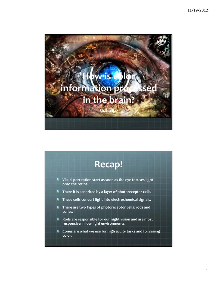
How is color information processed in the brain?
Shahn Ijaz
Recap!
Visual perception start as soon as the eye focuses light
- nto the retina.
There it is absorbed by a layer of photoreceptor cells. These cells convert light into electrochemical signals. There are two types of photoreceptor cells: rods and cones. Rods are responsible for our night vision and are most responsive in low light environments. Cones are what we use for high acuity tasks and for seeing color.