Gabriel Kreiman Email : gabriel.kreiman@tch.harvard.edu Phone : - - PowerPoint PPT Presentation

gabriel kreiman email
SMART_READER_LITE
LIVE PREVIEW

Gabriel Kreiman Email : gabriel.kreiman@tch.harvard.edu Phone : - - PowerPoint PPT Presentation

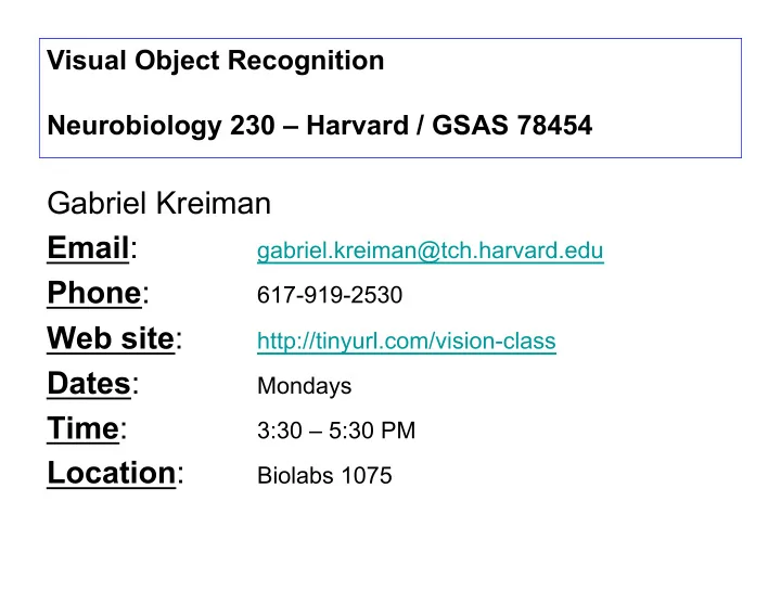
Visual Object Recognition Neurobiology 230 Harvard / GSAS 78454 Gabriel Kreiman Email : gabriel.kreiman@tch.harvard.edu Phone : 617-919-2530 Web site : http://tinyurl.com/vision-class Dates : Mondays Time : 3:30 5:30 PM Location :


slide-1
SLIDE 1

Visual Object Recognition Neurobiology 230 – Harvard / GSAS 78454

Gabriel Kreiman Email: gabriel.kreiman@tch.harvard.edu Phone: 617-919-2530 Web site: http://tinyurl.com/vision-class Dates: Mondays Time: 3:30 – 5:30 PM Location: Biolabs 1075

slide-2
SLIDE 2

Starting from the very beginning

  • Objects reflect light
  • Light photons impinge on the retina
  • The retina conveys visual information to the brain

An oversimplified (and rather erroneous) first-order description: The retina functions as a very sophisticated and spectacular digital camera

slide-3
SLIDE 3

Natural images are special

We only encounter a small subset of the space of possible images

  • Consider a grayscale image with 256 possible tones
  • Consider an image of size 100 x 100 pixels
  • How many such images are possible?

Answer For a size of 1x1 pixel, there are 256 possible images. For a size of 1x2 pixels, there are 2562 possible images. For a size of 100x100 pixels, there are 25610000 possibilities*.

*Some of those are “related” by translation or rotation or inversion, etc

Yet, we only encounter a small fraction of these possibilities in natural images

slide-4
SLIDE 4

Natural image statistics

Power spectrum ~ 1/f2

Simoncelli and Olshausen 2001

log( f (w)) = α log(w) + c w' → aw log( f (w')) = β log(w) + d

Note: Scale invariance There are multiple examples of power law distributions in physics, biology and social sciences

slide-5
SLIDE 5

Spatial aspects of natural scenes

The properties of nearby points are correlated

Simoncelli and Olshausen 2001

slide-6
SLIDE 6

Natural image statistics

There are also strong correlations in time The visual input is largely static, except for:

  • External object movements
  • Head movements
  • Eye movements

The visual image is largely static over hundreds of milliseconds

Silent Reading 225-250 ms fixation, 2 degrees saccade size (8-9 letters) Scene Perception 260-330 ms fixation, 4 degrees saccade “Slowness” has been proposed as a constraint for learning about objects (Foldiak 1991, Stringer et al 2006, Wiskott et al 2002, Li et al 2008)

slide-7
SLIDE 7

The image is focused onto the retina

slide-8
SLIDE 8

An image as a collection of pixels

57 53 58 63 44 41 66 93 68 25 67 33 52 117 130 121 124 119 130 94 34 58 65 106 67 71 84 152 164 142 150 145 143 111 64 47 55 98 104 117 124 130 147 147 79 44 40 67 89 80 78 91 107 97 87 68 44 51 60 66 61 61 69 66 52 48 47 79 99 57 47 44 47 54 46 41 41 50 110 123 70 44 46 45 51 49 43 40 61 87 95 58 45 55 46 46 51 49 39 62 72 87 63 59 59 57 48 56 47 44 49 51 52 52 52 48 48 51 52 55 56

slide-9
SLIDE 9

The retina

A beautiful circuitry composed of many different cell types

Dowling (2007), Scholarpedia, 2:3487 Wandell (1995), Foundations of Vision. Sinauer Books

  • ~0.5 mm thick
  • 5 x 5 cm retinal area
  • Three cellular layers
  • Rods (low-illumination conditions, ~108)
  • Cones (high-sensitivity, ~ 106)
  • Blind spot
  • Fovea (rod free, ~0.5 mm, ~ 1.7 deg)
  • Midget ganglion cells (small dendritic arbors)
  • Parasol ganglion cells (large dendritic arbors)
slide-10
SLIDE 10

Rods see largely in grayscale

slide-11
SLIDE 11

The retina

Some cells fire action potentials whereas other cells show graded responses

John Dowling (2007), Scholarpedia, 2:3487.

  • Photoreceptors transduce incoming light

input into electrical signals

  • Rod to bipolar convergence increases rod-

pathway sensitivity

  • Cones, rods, horizontal and bipolar cells are

non-spiking neurons

  • Many different types of amacrine cells
  • Retinal ganglion cells fire action potentials

and carry the output signals

slide-12
SLIDE 12

There is much more detail at the fovea

slide-13
SLIDE 13

The receptive field

Neurons throughout the visual system are very picky about the stimulus location

Fixation point Spike responses Receptive field This cartoon neuron responds only when a flash of light appears in the periphery, in the lower left quadrant Blumberg and Kreiman, 2010

slide-14
SLIDE 14

Physiology of retinal ganglion cells

The receptive field of most RGCs has a center-surround structure

Kuffler, S. (1953)

  • J. Neurophys. 16: 37-68
slide-15
SLIDE 15

Diversity of retinal ganglion cells

Minority of RGCs have more complex response properties:

  • Phasic cells respond briefly to stimulus onset, offset, or both
  • Some phasic cells respond selectively to edge orientation
  • Suppressed-by-contrast cells fire except when an edge is present in receptive field
  • Bistratified RGCs lack surrounds and are color-sensitive
  • Color-opponent cells have centers and surrounds with opposing color preferences
  • Intrinsically photosensitive RGCs contain photoreceptors and project to regions

controlling pupil size, circadian rhythm, etc.

  • Direction-sensitive cells respond to direction of motion of light or dark spots

These cells likely account for approximately 10% of RGCs Unclear to what extent they contribute to visual object recognition

Stone and Fukuda, Journal of Neurophysiology 1974 Cleland and Levick, Journal of Neurophysiology 1974 Berson et al., Science 2002

slide-16
SLIDE 16

The lateral geniculate nucleus

D(x,y) = ± 1 2πσcen

2 exp − x2 + y2

2σcen

2

% & ' ( ) * − B 2πσsur

2 exp − x2 + y2

2σsur

2

% & ' ( ) * + ,

  • .

/ D(x,y,t) = ± Dcen(t) 2πσcen

2 exp − x2 + y2

2σcen

2

% & ' ( ) * − BDsur(t) 2πσsur

2

exp − x2 + y2 2σsur

2

% & ' ( ) * + ,

  • .

/ Dcen(t) = αcen

2 texp −αcent

[ ]− βcen

2 texp −βcent

[ ]

Dsur(t) = αsur

2 texp −αsurt

[ ]− βsur

2 texp −βsurt

[ ]

Dynamic receptive fields in the retina/LGN

Dayan and Abbott. (2001) Theoretical Neuroscience. The MIT Press

slide-17
SLIDE 17

Difference of Gaussians

The center-surround structure can be described by a difference of gaussians (mexican-hat)

D(x,y) = ± 1 2πσcen

2 exp − x2 + y2

2σcen

2

% & ' ( ) * − B 2πσsur

2 exp − x2 + y2

2σsur

2

% & ' ( ) * + ,

  • .

/

Dayan and Abbott. (2001) Theoretical Neuroscience. The MIT Press

Center response (σcen) Surround response (σsur)

slide-18
SLIDE 18

Difference of Gaussians

The center-surround structure can be described by a difference of gaussians (mexican-hat)

Dayan and Abbott. (2001) Theoretical Neuroscience. The MIT Press

slide-19
SLIDE 19

To cortex, through the thalamus

The lateral geniculate nucleus (LGN) is the main visual part

  • f the thalamus:
  • 6 layers
  • Layers 2, 3 and 5 receive ipsilateral input
  • Layers 1, 4 and 6 receive contralateral input
  • Layers 1-2: magnocellular cells that receive input from M

ganglion cells

  • Layers 3-6: parvocelluar cells that receive input from P

ganglion cells

  • Between the layers: koniocellular cells that receive input

from bistratified retinal ganglion cells

  • Right and left visual hemifields are separate in the LGN
  • Right and left eyes are separate in the LGN
  • The visual field is represented multiple times in the LGN
  • On and Off center cells are present in all layers
  • LGN does not project back to the retina

NOTE: Most of the input to the LGN comes from visual cortex and not from the retina! (e.g. Douglas and Martin 2004) Wandell (1995), Foundations of Vision. Sinauer Books

slide-20
SLIDE 20

Subcortical visual pathways

Retinal projections Lateral geniculate nucleus (LGN) – Thalamus Superior Colliculi – Main visual pathway in birds, reptiles, fish Implicated in saccade generation in mammals Suprachiasmatic Nucleus – Hypothalamus: involved in circadian rhythms Pretectum

Pregeniculate Accesory optic system

Primates can recognize objects after lesions to the Superior Colliculus but not after lesions to V1 (Gross 1994 for historical overview).

slide-21
SLIDE 21

Visual system circuitry

Felleman and Van Essen. Cerebral Cortex 1991

slide-22
SLIDE 22

Further reading

Further reading

  • Class notes: http://tinyurl.com/vision-class
  • Wandell B. Foundations of Vision. Sinauer Books1995.
  • Dayan and Abbott. Theoretical Neuroscience. MIT Press 2001.

Some of the original articles cited in class (see lecture notes for full list)

  • Simoncelli and Olshausen. Annual Review of Neuroscience 2001
  • Dowling J. Scholarpedia 2007.
  • Felleman and Van Essen. Cerebral Cortex 1991.
  • Blumberg and Kreiman. Journal of Clinical Investigation 2010.
  • Kuffler. Journal of Neurophysiology 1953.
  • Foldiak. Neural Computation 1991.