from Single Molecules to Pathways Zn,Cu-SOD IMS Cu + Cu + Cox11 - - PowerPoint PPT Presentation

from single molecules to pathways
SMART_READER_LITE
LIVE PREVIEW

from Single Molecules to Pathways Zn,Cu-SOD IMS Cu + Cu + Cox11 - - PowerPoint PPT Presentation

Recent Advances in Biomolecular NMR Cytoplasm MT Zn,Cu-SOD GSH NMR in Cellular Structural Biology: Mitochondrion CCS CCS from Single Molecules to Pathways Zn,Cu-SOD IMS Cu + Cu + Cox11 MT Matrix CCO Cox17 2S-S Ctr1/2 HAH1 Lucia


slide-1
SLIDE 1

Zn,Cu-SOD

HAH1

MT

GSH

Ctr1/2 CCS

Cu+

ATP7A/B

Mitochondrion

Cox17

D1 D2 D3 D4 D5 D6

Golgi complex

Cu+

Zn,Cu-SOD

CCS

MT

IMS

GSH

Sco2 Matrix

Cox11

Cytoplasm

CCO Sco1

Cox172S-S SecPr SecPr

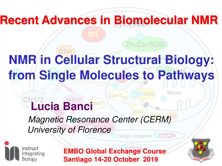
NMR in Cellular Structural Biology: from Single Molecules to Pathways

Lucia Banci

Magnetic Resonance Center (CERM) University of Florence

Recent Advances in Biomolecular NMR

EMBO Global Exchange Course Santiago 14-20 October 2019

slide-2
SLIDE 2

Molecular biology, cellular biology and biophysics labs

GENEXPRESS

Genetic expression laboratory

*equipped with cryo probes

Library Workstations

ss700wb 600 500* 400 700* ss850wb 950* 1200** 900*

He Liquifier Relaxometer

(0.01-40 MHz)

600

CERM/CIRMMP a core center of Instruct

Competence Center

ss800

Cryo EM**

** due in 2019

400 700* 700*

Q,X-band pulsed EPR Biobank Mass Spectrometry X-Ray Crystallography

11 NMR spectrometers + 1 Relaxometer, The largest available magnetic field range (0.01 – 950 MHz)……soon up to 1200 MHz Conference Room Access available through INSTRUCT-ERIC and iNEXT-Discovery

slide-3
SLIDE 3

The Cermians

slide-4
SLIDE 4

Recent Advances in Biomolecular NMR

  • Cellular Structural Biology

To describe and understand biological processes at molecular level

  • In cell NMR

For studying biomolecules in a cellular context

  • Structural Vaccinlogy

Rational vaccine design based on the structural knowldge of the antigene

slide-5
SLIDE 5

Living systems are complex: mixture of proteins, nucleic acids, other biomolecules, several cellular compartments,...etc

A Systems Biology approach is needed. All the players involved in a given process have to be considered as well as their 3D structural and dynamical interactions determined.

Proteins must be framed within their cellular context

Integrating a Cellular Approach with Atomic Resolution

slide-6
SLIDE 6

Integrating Atomic Resolution with the Cellular Context

No free copper ions in the cytoplasm

Copper trafficking in human cells

Nucleus Golgi Ctr

Regulators MNK/WLN

Amine Oxidase, Lysyl oxidase

Cu(I)

?

Cu(II) Cu(I)

Cox23 Cox23 Cox19Cox19

Mitocondria

Sco1 Sco2 Cox11 MT ? CCS SOD CCO

Ceruloplasmin

SOD1 CCS Hah1 MT Cox17 Cox17

GSH GSSG E° of cytosolic glutathione = -289 mV, corresponding to GSH and GSSG in vivo concentrations of 13 mM and 0.7 mM

slide-7
SLIDE 7

Let’s start with a single process

monomeric apo hSOD1SH-SH

Copper binding

C57 C146

Zinc binding Disulfide bond formation Zn Cu SS bond

These post translational modifications affect the fold properties and monomer/dimer equilibrium

Cu Zn

dimeric (Cu2,Zn2) hSOD1SS Active enzyme: (2O2

  • + 2H+

O2+ H2O2)

SOD1: present in cytoplasm, mitochondrial IMS, nucleus, peroxisomes

Maturation of Cu,Zn-SOD1

slide-8
SLIDE 8

In-cell NMR can monitor functional processes in live human cells

Isotopically labelled proteins are overexpressed and directly

  • bserved by hi-res NMR in

living human cells. Transfected HEK293T cells are used as a model system for human cells Maturation processes such as protein folding, post translational modifications (i.e. metal binding, disulfide bond formation) are followed at atomic resolution. Understanding intracellular processes at the molecular level requires a high resolution description. In-cell NMR provides atomic-level information on a protein in the cellular environment.

HS SH

1H

15N

Cys 146 Cys 111 Cys 6 SH Cys 57

1H 15N

apo-SOD1 E,Zn-SOD1 apo-SOD1

slide-9
SLIDE 9

In-cell NMR can monitor functional processes in live human cells

Isotopically labelled proteins are overexpressed and directly

  • bserved by hi-res NMR in

living human cells. Transfected HEK293T cells are used as a model system for human cells Maturation processes such as protein folding, post translational modifications (i.e. metal binding, disulfide bond formation) are followed at atomic resolution. Understanding intracellular processes at the molecular level requires a high resolution description. In-cell NMR provides atomic-level information on a protein in the cellular environment.

+ Zn(II)

HS SH HS SH SH HS

1H

15N

Cys 146 Cys 111 Cys 6 SH Cys 57

1H 15N

E,Zn-SOD1 E,Zn-SOD1

slide-10
SLIDE 10

In-cell NMR can monitor functional processes in live human cells

Isotopically labelled proteins are overexpressed and directly

  • bserved by hi-res NMR in

living human cells. Transfected HEK293T cells are used as a model system for human cells Maturation processes such as protein folding, post translational modifications (i.e. metal binding, disulfide bond formation) are followed at atomic resolution. Understanding intracellular processes at the molecular level requires a high resolution description. In-cell NMR provides atomic-level information on a protein in the cellular environment.

SH SH S S S S

+ Cu(I) + Zn(II)

HS SH HS SH SH HS

1H

15N

Cys 146 Cys 111 Cys 6 SH S-S Cys 57

1H 15N

Cu,Zn-SOD1 Cu,Zn-SOD1

Banci L, Barbieri L, Bertini I, Luchinat E, Secci E, Zhao Y, Aricescu AR, Nat Chem Biol, 2013

slide-11
SLIDE 11

HS SH SH HS HSSH SH SH SH SH HS SH SH HS SH SH HS HS HSSH SH HS

Zn(II)

S S S S S S S S HS HS

CCS

SH SH S S S S

Cu(II) Zn(II) Cu(I)

Following SOD1 maturation steps in human cells

E,E-SOD1SH E,Zn-SOD1SH E,Zn-SOD1S-S Cu(I),Zn-SOD1S-S

Banci L, Barbieri L, Bertini I, Luchinat E, Secci E, Zhao Y, Aricescu AR, Nat Chem Biol, 2013

slide-12
SLIDE 12

Incomplete maturation of SOD1 fALS mutants

  • ALS: a motor neuron disease
  • 20% of familial cases is related

to mutations of SOD1.

  • 165 mutations identified so far,

scattered throughout the sequence.

  • Mutations are thought to cause defects in SOD1 maturation,

promoting aggregation of the apo protein

Luchinat E, Barbieri L, Rubino JT, Kozyreva T, Cantini F, Banci L, Nat. Comm., 2014

slide-13
SLIDE 13

Maturation defects of fALS SOD1 mutants

Luchinat E, Barbieri L, Rubino JT, Kozyreva T, Cantini F, Banci L, Nat. Comm., 2014

Many SOD1 mutants do not bind zinc in the cell, and accumulate as an unstructured species, which does NOT evolve toward the native form This unstructured species DOES NOT bind zinc It could be a precursor of SOD1 aggregates

(A4V, I35T, G37R, G85R, G93A, I113T) I113T G93A

I113T#

I113T

The mutations do not affect zinc binding in vitro

slide-14
SLIDE 14

Copper trafficking in human cells

Nucleus Golgi

Hah1

Ctr

Regulators MNK/WLN

Amine Oxidase, Lysyl oxidase

CCS

Cu(I)

?

Cu(II) Cu(I)

Cox17 SOD1 MT Cox23 Cox23 Cox19 Cox19

Mitocondria

Sco1 Sco2 Cox11 MT ? CCS SOD CCO

Ceruloplasmin

No free copper ions in the cytoplasm

slide-15
SLIDE 15

Banci, Bertini, Ciofi-Baffoni, Karit, Kozyreva, Palumaa, Nature, 2010

Towards systems biology of copper

The knowledge of the structures of the proteins and of their complexes allows the atomic level description of the transfer processes

slide-16
SLIDE 16

System-wide understanding

  • f biological processes requires an

atomic-level description of the functional processes

slide-17
SLIDE 17

Iron Sulfur Biogenesis in human cells

Ciofi-Baffoni S, Nasta V, Banci L., Metallomics. 2018, 49-72 Humans have around 70 Fe-S proteins! They are less than 0.5% of the human proteome, but absolutely essential. And the Fe-S clusters need to be synthesized!

slide-18
SLIDE 18

Structural properties of Anamorsin An essential protein for FeS cluster biosynthesis

Mia40 recognition site Intrinsically Disordered Domain N-terminal domain Folded domain “standard” approach Linker [2Fe-2S] cluster Highly paramagnetic with fast relaxation and no PCS The worst case!! Mia40 forms two disulfide bonds when in mitochondria This motif binds a cluster in the cytoplasm

slide-19
SLIDE 19

Playing For or Against Paramagnetic Relaxation?

Exploit the differences in longitudinal relaxation

30 ms < 6-10 Å T1 < 100 ms 100 ms < 10-11 Å T1< 200 ms 500 ms

Inversion Recovery

The effect of T1 relaxation 200 ms 100 100 ms ms 50 50 ms ms 20 ms 10 ms 2 ms IR delays selected according to T1 values 3-6 Å T1< 30ms

IR is combined with fast recycling times

slide-20
SLIDE 20

0.25 0.5 0.75 1 0.002 0.004 0.006 t(s)

no relax

100ms 10 ms 5 ms 2 ms 1 ms 0.5 ms

Playing For or Against Paramagnetic Relaxation?

Limit the negative effects of fast transverse relaxation

The effect of T2 relaxation

INEPT coherence transfer

We can detect peaks for signals with Dn up to ca. 300 Hz

ppm

d 13C Remove the reverse INEPT Phasing AP in dispersion mode

Antiphase signal acquisition

slide-21
SLIDE 21

Tailored 15N HSQC of the [2Fe-2S]-domain of anamorsin

1H 15N 15N- IR-HSQC-AP 15N 15N- HSQC 1H

13 HN peaks, missing in standard 15N HSQC, can be detected.

1H T1 values range from 5 to 30 ms

Banci et al PNAS 2013, 2014; Ciofi-Baffoni, Gallo, Piccioli, J. Biomol NMR 2014

slide-22
SLIDE 22

Paramagnetic-tailored 13C-direct CACO of [2Fe-2S]- domain of anamorsin

13C 13C 13C 13C 13C-CACO-IPAP 13C-CACO-AP

T250 G252 S236 K244

13C signals, absent in standard 13C-direct esperiments, are also observed

via 13C COSY and tailored CON Overall, about 10 additional 13C resonances are detected

slide-23
SLIDE 23

Structure of the [2Fe-2S] cluster binding region in anamorsin

Blue - residues detected in the “diamagnetic” experiments Cyano - residues whose 13C or 15N signals were detected in paramagnetic-tailored 13C or 15N experiments

T250 K244 G252 S236

The “paramagnetic” 13C and 1H T1s provide structural info around the FeS cluster

The overall structure was then subjected to MD trajectory in explicit water

slide-24
SLIDE 24

Banci, Ciofi-Baffoni, Gajda, Muzzioli, Peruzzini, Winkelmann, Nat. Chem Biol. 2015

No transfer

The Trx domain of GRX3 is essential for protein-protein recognition

Anamorsin receives the [2Fe-2S] clusters from GRX3 in the cytoplasm

Cluster site characterization complemented with EPR and Moessbauer data

slide-25
SLIDE 25

…but in oxidative conditions, the BolA2-GRX3 complex is more efficient in anamorsin maturation

Banci L, Camponeschi F, Ciofi-Baffoni S, Muzzioli R. J Am Chem Soc. 2015, Nuttle, X. et al. Nature 2016

The GRX3/BolA2 heterocomplex is more stable in oxidative conditions An alternative route to anamorsin maturation

anamorsin

slide-26
SLIDE 26

Electron transfer between Ndor1 and anamorsin

Anamorsin tightly interacts with Ndor1 through its flexible, unstructured linker The [2Fe-2S]-CIAPIN1 domain transiently interacts with the FMN domain

  • f Ndor1 transferring one electron from FMN to the [2Fe-2S] cluster.

Anamorsin Ndor1 (closed conformation) Ndor1 (open conformation) Anamorsin Anamorsin

NADPH FAD FMN

[2Fe-2S]- CIAPIN1 domain

Banci , Bertini, Calderone, Ciofi-Baffoni, Giachetti, Jaiswal, Mikolajczyk, Piccioli, Winkelmann PNAS, 2013

FMN-binding domain of Ndor1 Flexible linker N-terminal domain

FMNH2

slide-27
SLIDE 27

Electron transfer between Ndor1 and anamorsin

Banci , Bertini, Calderone, Ciofi-Baffoni, Giachetti, Jaiswal, Mikolajczyk, Piccioli, Winkelmann PNAS, 2013

slide-28
SLIDE 28

Iron Sulfur Biogenesis in human cells

Ciofi-Baffoni S, Nasta V, Banci L., Metallomics. 2018, 49-72

slide-29
SLIDE 29

Brancaccio, Gallo , Mikolajczyk, Zovo, Palumaa, Novellino, Piccioli, Ciofi-Baffoni, Banci JACS 2014

Iron Sulfur Biogenesis in Mitochondria Cluster transfer between GRX5 and ISCA1/2

Apo ISCA1/ISCA2 [2Fe-2S] GRX5a [2Fe-2S] GRX5b

+

[4Fe-4S] ISCA1/ISCA2 apo GRX5

Ser41 Ser41 Cys38 Cys38

Combining “standard” NMR, paramagnetic NMR, EPR, mass spec, etc….we learned how GRX5 transfers the cluters to ISCA1/ISCA2 and the 4Fe4S cluster is formed.

Banci, Ciofi-Baffoni, Del Conte, Gadepalli, Neri, Piccioli, Winkelmann PNAS 2014

GSH

slide-30
SLIDE 30

WT apo ISCA2 WT [4Fe-4S] ISCA2

 Cys 79 is essential for binding a [2Fe-2S] or a [4Fe-4S] cluster  Cys 144 and Cys 146 provide the other two binding residues  Binding of a [2Fe-2S] cluster can occur with Cys 79, and either Cys 144 or Cys 146, without the need of all three conserved Cys of ISCAs

WT [4Fe-4S] ISCA2

Fe/S cluster binding properties of ISCAs

Brancaccio D, Gallo A, Piccioli M, Novellino E, Ciofi-Baffoni S, Banci L., JACS, 2017

slide-31
SLIDE 31

N-terminus C-terminus b1 b3 b2 a1 a2 h2 h1 a4 H67 a3 H102

BOLA1

H58 H86 N-terminus C-terminus H96 b2 C59 b1 b3 a1 h1 a2 h2

BOLA3

H81

NMR solution structures of human BOLA1 and BOLA3

H58 H67 H102 H96 C59 H86 H81

BOLA1 BOLA3

PDB ID 5LCI PDB ID 2NCL Uzarska MA, Nasta V, Weiler BD, Spantgar F, Ciofi-Baffoni S, Saviello MR, Gonnelli L, Mühlenhoff U, Banci L, Lill R. Elife, 2016

BOLAs alone do not bind FeS clusters but they do when complexed with GRX5

slide-32
SLIDE 32

Structural basis of the interaction between human apo- and holo-GRX5 with the BOLA proteins

Uzarska M, Nasta V, Weiler B, Spantgar F, Ciofi-Baffoni S, Saviello M, Gonnelli L, Mühlenhoff U, Banci L, Lill R. Elife, 2016

Apo interaction surfaces Holo interaction surfaces Cluster ligands were identified through mutations and para NMR and EPR data Nasta, Giachetti, Ciofi-Baffoni, Banci Biochim Biophys Acta – General Subjects 2017

slide-33
SLIDE 33

GRX5 BOLA3

[2Fe-2S]2+ C59 C67 GSH H96 H67 GSH C67 H102 [2Fe-2S]1+

BOLA1 GRX5

Electron transfer Cluster transfer

Solvent exposed cluster Buried cluster

The structures of [2Fe-2S] GRX5-BOLAs support two different functions for the two heterocomplexes

Nasta, Giachetti, Ciofi-Baffoni, Banci Biochim Biophys Acta – General Subjects 2017

slide-34
SLIDE 34

Iron Sulfur Biogenesis in human cells

Ciofi-Baffoni S, Nasta V, Banci L., Metallomics. 2018, 49-72

slide-35
SLIDE 35

An integrated approach to investigate electrons and FeS cluster transfer processes

ESI-MS Para-NMR Solution NMR, X-ray, Protein docking Biophysics: EPR, UV-vis, Mössbauer CD …

Banci et al. PNAS 2014 Banci, Ciofi-Baffoni, Bill et al. JBIC 2013 Banci, Ciofi-Baffoni et al. PNAS 2013 Banci, Ciofi-Baffoni et al. Chem Biol. 2011

Eukaryotic Fe/S assembly machineries

FF parameters determination

slide-36
SLIDE 36

Structural Vaccinology: the structure-based rational vaccine design

slide-37
SLIDE 37

vaccine

STRUCTURAL VACCINOLOGY

Cultivate microorganism Genome-based approaches Pan-genome approach Determination of antigens structure

past present

slide-38
SLIDE 38
  • 900 MHz Spectrometer (298K)

Chemical shift mapping

1H-15N HSQC-TROSY

  • f fHbpC alone

1H-15N HSQC-TROSY

  • f fHbpC:mAb502

mixture

Interaction between fHbpC and a fAb portion

  • f the antibody mAb502 (as studied by NMR)

The data show that mAb502 recognizes a conformational epitope within a well-defined area of the immunodominant C-terminal domain of fHbp.

1H-15N HSQC-TROSY of

fHbpC alone

1H-15N HSQC-TROSY-

CRINEPT

  • f fHbpC:mAb502 mixture

Residues predicted by immunological data and confirmed by NMR NMR data suggested the involvement of

  • ther amino acids
slide-39
SLIDE 39
  • 1. They are still solvent accessibles!
  • 2. fHbpC contains the major part of the epitope

Model of the complex between fHbpC & fAb portion of mAb502

Residues of fHbpC involved in binding to mAb502 mapped onto the full length protein structure

slide-40
SLIDE 40

Structure of antigen fHbp Heavy chain of monoclonal antibody Mab502 Light chain of monoclonal antibody Mab502 Fab region

  • f

antibody

Complex of a monoclonal antibody with a Meningococcus B antigen (Factor H binding protein)

Scarselli, Cantini, Banci, Rappuoli et al., Science Transl. Med. 2011 fHbp is very effective in inducing protective immunity eliciting antibodies but has different sequence in different strains of MenB

slide-41
SLIDE 41

Structure-based design of a vaccine against Mengingococcus B

Scarselli, Cantini, Banci, Rappuoli et al., Science Transl. Med. 2011 By knowing the structural properties

  • f the antigen and of the epitopes in

all the variants, a chimera antigen was produced which elicits complete protective immunity

slide-42
SLIDE 42

1D 1H NMR spectra at various fields

1.2 mM α-synuclein in 20 mM KPi, 100 mM NaCl, 50 μM EDTA, pH 6.5, 283 K 120 µl sample in 3 mm shighemi tube CP TCI probehead

1.2 GHz

d1H/ppm

The first spectra at 1.2 GHz

slide-43
SLIDE 43

fixed Hz/cm

1D 1H NMR spectra at various fields

slide-44
SLIDE 44

1H-15N HSQC at 1.2 GHz

1.2 mM α-synuclein sample in 20 mM KPi, 100 mM NaCl, 50 μM EDTA, pH 6.5

500 MHz 700 MHz 950 MHz 1.2 GHz

slide-45
SLIDE 45

proline fingerprint

2D CON spectrum at 1.2 GHz

207 AA 140 AA α-synuclein CBP-ID4

slide-46
SLIDE 46

1.2 mM α-synuclein sample in 20 mM KPi, 100 mM NaCl, 50 μM EDTA, pH 6.5

Signals linewidth at half-height is about 5 Hz

15N-detected TROSY spectrum at 1.2 GHz

slide-47
SLIDE 47

~1 mM α-synuclein sample in 20 mM KPi, 100 mM NaCl, 50 μM EDTA, pH 6.5

15N-1H TROSY spectrum at 1.2 GHz

BLUE: TROSY CYAN: TROSY with Ha-decoupling in F2 Linewidth in 15N TROSY:  = 5.0 Hz

  • w. dec:  = 3.7 Hz

Linewidth in 1H  = 17.0 Hz

slide-48
SLIDE 48

Thank you for your attention !!

slide-49
SLIDE 49

Structural and biophysical methods for biological macromolecules in solution

EMBO Global Exchange Lecture Course 14 – 20 October 2019 | Santiago, Chile