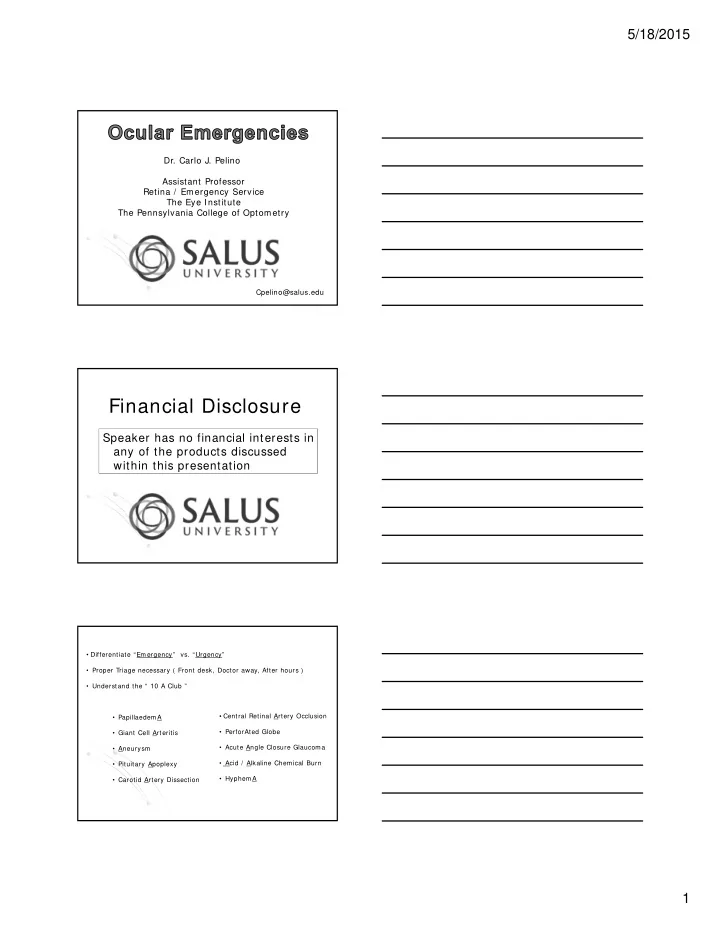
financial disclosure
play

Financial Disclosure Speaker has no financial interests in any of - PDF document

5/18/2015 Dr. Carlo J. Pelino Assistant Professor Retina / Emergency Service The Eye Institute The Pennsylvania College of Optometry Cpelino@salus.edu Financial Disclosure Speaker has no financial interests in any of the products discussed


  1. 5/18/2015 Dr. Carlo J. Pelino Assistant Professor Retina / Emergency Service The Eye Institute The Pennsylvania College of Optometry Cpelino@salus.edu Financial Disclosure Speaker has no financial interests in any of the products discussed within this presentation • Differentiate “Emergency” vs. “Urgency” • Proper Triage necessary ( Front desk, Doctor away, After hours ) • Understand the “ 10 A Club ” • Central Retinal Artery Occlusion • PapillaedemA • PerforAted Globe • Giant Cell Arteritis • Acute Angle Closure Glaucoma • Aneurysm • Acid / Alkaline Chemical Burn • Pituitary Apoplexy • HyphemA • Carotid Artery Dissection 1

  2. 5/18/2015 Corneal Abrasion Corneal Ulcer CRAO CRAO Cherry Red Spo t Cilioretinal artery sparing 2

  3. 5/18/2015 • Rapid unilateral painless vision loss – count fingers / light perception visual acuity in 90 % of eyes Etiology: • Arterial emboli ( cholesterol, fibro-platelet, calcific ) 20% cases • Thrombus formation secondary to arteriosclerosis • Arteritis – Giant cell arteritis, Lupus, Polyarteritis nodosa • Blood dyscrasias (hypercoagulation disorder) • Migraine • Optic nerve drusen Presentation: • Superficial retinal whitening with a “cherry red spot” in the fovea • Retinal whitening within 30 minutes to 1 hour from ischemic necrosis • Narrowed retinal arterioles with segmentation • Cilioretinal artery may be spare the fovea • Relative Afferent Pupillary Defect • Optic Nerve pallor weeks to months later Treatment: • Ocular Massage (digital pressure) – 10 sec on/ 10 sec off x 5 minutes • Vasodialtion (increase CO2 by breathing into a paper bag) • Lowering of intraocular pressure(IOP) … . beta blocker, Diamox, etc. • Anterior chamber paracentesis • Sublingual Nitroglycerin / Intra-Arterial fibrinolysis with ( tPA ) Systemic Testing: • Immediate Westergren Sed. Rate if patient is > 55 years old; R/ O GCA • Carotid artery evaluation, blood pressure, lipid profile, blood sugar • Echocardiogram – cardiac evaluation • Fluorescein angiography (FA) 3

  4. 5/18/2015 Work-Up: • Cardiovascular Evaluation Echocardiogram (Transesophageal vs.Transthroacic) To find congenital / acquired valvular anomalies To evaluate for cardiac myxoma, atrial fibrillation Congenital heart defects such as a patent foramen ovale R/ O endocarditis (infectious process)-Heart murmur 90% • Hyperviscosity Evaluation Multiple Myeloma, leukemias, and Waldenstroms Macroglobulenemia • Hypercoaguable Evaluation Factor V Leiden, Hyperhomocysteine, Antiphospholipid syndrome, birth control pills, pregnancy • Hemoglobinopathies Sickle Cell disease and trait Work Up: • Rule out Vasculitis: Systemic Lupus Erythematosus, polyarteritis nodosa • Rule out Atherosclerotic risk factors: Diabetes, hypertension, hyperlipidemia and smoking • Carotid Artery Evaluation: Ipsilateral carotid artery at its bifurcation Intracranial internal carotid artery siphon BRAO patients should undergo a thorough medical evaluation BRAO / CRAO may be the first warning sign of a serious disorder Workup should be tailored to the age of the patient – Life expectancy for patients with CRAO is 5.5 years vs. 15.4 years for an age matched population without CRAO Follow Up: • RTC at 1 month to check for neovascularization of disc/ iris • RTC at 3 months to check for neovascularization of disc/ iris • Neo of iris = 20 % of patients at about 4 weeks • Neo of disc = 3 % of patients • Extremely important to perform a complete medical work-up to stop progression of the disease along with any systemic sequelae 4

  5. 5/18/2015 • Must be differentiated from “Non-Arteritic” AION • Sudden painless loss of vision • Females > Males ( 2: 1 ratio ) • Patients usually > 55 years of age NAION AION • Etiology Occlusion of the “short posterior ciliary” arteries with giant white blood cells Short Posterior Ciliary Arteries Presentation: Ocular “Giant Cell Arteritis” • Acute painless vision loss (VA loss is usually permanent) • Pale swelling of the optic nerve head with flame shaped hemes • Central retinal artery occlusion may occur • Cranial nerve palsy (CN 3,4,6) may also be present, CWS Presentation: Systemic Arteritic AION • Headache • Scalp tenderness • Jaw claudication • Night sweats • Weight loss • Fever • Polymyalgia rheumatica • Depression Laboratory Testing: • Immediate Erythrocyte Sedimentation Rate (Westergren ESR) • Immediate C –reactive protein ( Acute Phase Reactant ) >2.45 mg/dl • Platelet count ( Thrombocytosis ) = risk for permanent visual loss • CBC with differential = anemia of chronic inflammation 5

  6. 5/18/2015 • A normal ESR does not R/O GCA ; Normal in ~ 13 % of GCA • 20 % of GCA patients do not have systemic symptoms Giant Cell Arteritis White Blood Cells • Temporal Arteritis • T‐cells infiltrate arteries • Cranial Arteritis • Cytokines IL,TNF, IFN • Granulomatous Arteritis Attacks medium and large sized arteries • Superficial Temporal Artery • Coronary Artery • Subclavian Artery • Facial Artery Possible association with “Polymyalgia Rheumatica” (PMR) • Stiffness in the neck, shoulder, and hip • 50 % of Giant Cell patients have PMR • Is there a link between GCA and PMR ??? Treatment: • Immediate IV steroid therapy – IVMP x 3 days Oral Prednisone x 24 months • Possible temporal artery biopsy • Done within a week of starting steroids • Specimen is 2.5 cm long • If biopsy is negative but suspicion high, then biopsy the opposite side • ~ 13 % of cases will be positive on the opposite side • Methotrexate with Prednisone • Always suspect recurrence 6

  7. 5/18/2015 The “One Third Rule” in Giant Cell Arteritis • 1/3 of optic nerves in the fellow eye will become infarcted within 48 hours in untreated patients. • 1/3 of optic nerves in the fellow eye will become infarcted within 1 month in untreated patients. • Second eye infarctions are rare after more than 1 month “Malignant Hypertension” defined as Blood Pressure > 210/120 Ocular Presentation: General Rule: • Disc edema with or without exudate • Chest Pain • Arterio‐venous crossing changes • Difficulty breathing • Nerve fiber layer infarcts (cotton wool spots) • Immediate Attention • Macular edema • Hard exudates / flame shaped hemes • Choroidal ischemia Treatment: • Blood pressure measurement • Immediate referral to emergency room or PCP for slow lowering of the the blood pressure Grade 4 HR Grade 4 HR BP 225 / 125 BP 235 / 130 7

  8. 5/18/2015 “Malignant Hypertension” Hypertensive Choroidopathy Hypertensive Choroidopathy 8

  9. 5/18/2015 Orbital Cellulitis • Inflammation of orbital soft tissue posterior to the orbital septum • May get a direct extension to the brain – may lead to death Systems of Classification • Stage 1 Preseptal Cellulitis • Stage 2 Orbital Cellulitis • Stage 3 Subperiosteal abscess • Stage 4 Intraorbital abscess • Stage 5 Cavernous Sinus Thrombosis – usually has fatal outcome Etiology: • Eyelid infection (Hordeolum) • Sinus infection (paranasal sinusitis extension in~ 90% of cases) • Dental infection • Ocular trauma or surgery • Orbital infection (dacryocystitis) • Upper respiratory infection • Otitis Media Presentation Ocular: • Eyelid edema (absence of a lid crease) - Painful • Conjunctival chemosis • Proptosis / Globe displacement • Restricted motility- may have associated pain (60 % ) • Visual Acuity decrease Sinusitis • S . pneumoniae • other streptococci • S. aureus • H. influenzae (esp. in children) • anaerobes less common Post-traumatic and post surgical • S. aureus most common • anaerobes less common 9

  10. 5/18/2015 Usually, orbital cellulitis occurs in the childhood years which has been attributed to the relatively incomplete development of immunity in this age group. In these patients, sinus disease has been found to be the most common predisposing factor. Over 90% of these patients have radiologically confirmed sinusitis, the most common being ethmoidal and maxillary. Ethmoidal sinusitis has been demonstrated to be the source of infection in significantly large number of cases. Hordeolum Preseptal Cellulitis 10

  11. 5/18/2015 Presentation: Systemic Preseptal Cellulitis Lab Work • Fever ( as high as 104 ) • Complete Blood Count (CBC) • Headache • CT if unable to tell orbital involv. • Malaise • Nausea / Vomiting Treatment: • Immediate referral to emergency room or to Ophthalmology • Broad spectrum coverage with I V antibiotics after culture • MRI or orbital CT (Contrast not needed) • Hospitalization • Consultation with ENT, PCP and Neurosurgery if intracranial infection • Inflammation of orbital soft tissue posterior to the orbital septum Dacryocystis with Oral antibiotics Preseptal Cellulitis Orbital Cellulitis IV antibiotics Preseptal Cellulitis Oral antibiotics 11

Download Presentation
Download Policy: The content available on the website is offered to you 'AS IS' for your personal information and use only. It cannot be commercialized, licensed, or distributed on other websites without prior consent from the author. To download a presentation, simply click this link. If you encounter any difficulties during the download process, it's possible that the publisher has removed the file from their server.

Recommend


More recommend