
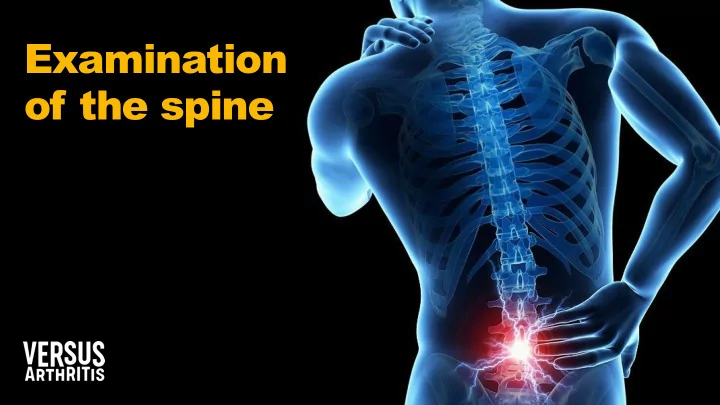
SLIDE 1
Examination
- f the spine
Examination of the spine Meet the speakers Dr Nadia Vawda Dr - - PowerPoint PPT Presentation
Examination of the spine Meet the speakers Dr Nadia Vawda Dr Christian Verrinder GP and Clinical Champion in Physical Activity with PHE GP with Special Interest in MSK Medicine Meet the speakers Dr Gile Dr Giles s Hazan Hazan Dr AN Dr
Dr Nadia Vawda
GP and Clinical Champion in Physical Activity with PHE
Dr Christian Verrinder
GP with Special Interest in MSK Medicine
Dr Gile Dr Giles s Hazan Hazan GPwSI GPwSI in MSK Medicine in MSK Medicine Dr AN Dr ANDRE DREW J W JACKSO CKSON GP with Special Interest in MSK Medicine Clinical lead VERSUS arthritis ‘core skills in msk’
history from an MSK patient, including eliciting red flags and psychosocial flags
the MSK patient
appropriate investigations, referral, safety net and follow-up
Compromise of roles between being upright and being able to bend and twist, so everyone gets it
consultations
1 in 3
adolescents experience lower back pain
Probably can’t prevent it Frequently recurs
50% of cases start with no
mechanism
(think about history)
Don’t over medicalise
Usually felt in the lumbar area (load) and cervical area (movement); increased clinical suspicion if thoracic pain is present Best predictor of malignancy:
Be alerted to other diagnoses if:
course
all shrink!)
is 30–50 years old; after this age, the risk decreases over time
may be a contributing factor
S1 level (i.e. below the knee dermatomes)
Von Korff M et al. (1996) The course of back pain in primary care. Spine (Phila Pa 1976). 21(24):2833–7.
Most people recover within approximately 6
WEEKS
Sciatica has a worse prognosis than LBP, with 30% of patients having clinically significant symptoms at 12 months
We can’t usually explain pain and prognosis by imaging
The prognosis for ‘disability’ is more dependent
than pathology; this can be predicted using the STarT tool, as per NICE guidelines People who are disabled by their back pain tend to worry too much about their back and/or not moving enough (what we say really matters) Positive messages:
mean harm
to be 100% to return to activity/work
conditions
neuropathic medications, injections
challenging yellow flags
✓ Exclude red flags ✓ Exclude inflammatory back pain ✓ Identify ‘nerve’ compression / pain vs. referred pain ✓ Stratify risk of disability (yellow flags) ✓ Manage the patient as per NICE guidelines
NICE (2016) Low back pain and sciatica in over 16s: assessment and management. [Accessed: 03/05/2019] Recommendation: 1.1.2. See appendix for full details.
Best predictor of malignancy:
Be alerted to other diagnoses if:
clinical course
Key red flags for identifying fractures are:
corticosteroids
contusion or abrasion Return
emergency
Developed by Dr Sue Greenhalgh, PhD, MA, GDPhys, FCSP, Professor Carole Truman
Leeds neurosurgical GP guidance on surgical indications.
with central cord compromise
improvement with conservative measures by six weeks (some improvement is likely to imply eventual resolution)
(>3 months)
Osteoarthritis Inflammatory Mechanical Persistent (chronic) pain or red flags
National Ankylosing Spondylitis Society.
1. Did your back pain start when you were aged <40?
and is likely to have onset below 45 years old
pain as opposed to only recording their current age as they may have been experiencing back pain for several years 2. Did your back pain develop gradually?
frequently of a more sudden onset. IBP has an insidious onset and patients are likely to have been experiencing back pain for >3 months 3. Does your back pain improve with movement?
with movement and exercise 4. Do you find there is NO improvement in your back pain with rest?
feature of inflammatory back pain 5. Do you suffer with back pain at night that improves upon getting up?
when resting at night with waking and getting up during the 2nd half of the night
✓ Exclude red flags ✓ Exclude inflammatory back pain ✓ Identify ‘nerve’ compression / pain vs. referred pain ✓ Stratify risk of disability (yellow flags) ✓ Manage the patient as per NICE guidelines
NICE (2016) Low back pain and sciatica in over 16s: assessment and management. [Accessed: 03/05/2019] Recommendation: 1.1.2. See appendix for full details.
Is this sciatica?
Often starts with back pain that settles to be replaced by acute leg pain; may have had recurrent episodes
preceding years Pain generally radiates to foot or toes (L5/S1) Numbness and paraesthesia in the same distribution Nerve irritation signs: Valsalva/ cough/sneeze Reduced SLR/slump which reproduces leg pain Motor, sensory or reflex changes; limited to one nerve root
Lower limb dermatome x2 HYPERLINK https://netterimages.com/dermatomes-of-lower-limb-labeled-anatomy-atlas-5e- internal-medicineprimary-care-frank-h-netter-4808.html
lordosis, pelvic shift, scars/wasting/rash)
their back are working
then move to a sitting position
hip, LLD, Babinski, peripheral pulses as relevant
prostate
✓ Exclude red flags ✓ Exclude inflammatory back pain ✓ Identify ‘nerve’ compression / pain vs. referred pain ✓ Stratify risk of disability (yellow flags) ✓ Manage the patient as per NICE guidelines
NICE (2016) Low back pain and sciatica in over 16s: assessment and management. [Accessed: 03/05/2019] Recommendation: 1.1.2. See appendix for full details.
The Keele STarT Back Screening Tool is a brief validated tool designed to screen primary care patients with low back pain for prognostic indicators that are relevant to initial decision making It risk stratifies patients into 3 groups:
– Low risk: low risk of chronicity – Medium risk: mainly physical obstacles to recovery – High risk: additional psychological obstacles to recovery
Each group should be offered a different package of care and provision of this has shown to be cost effective for both the NHS (£35.49/patient) and society (£675/patient) It’s conclusions led it to being included in NICE guidelines and becoming part of the ‘national back pain strategy’
https://startback.hfac.keele.ac.uk/ Hill JC et al. (2008) Arthritis Rheum,59:632-41.
NICE (2016) Low back pain and sciatica in over 16s: assessment and management. [Accessed: 03/05/2019]; Recommendation: 1.1.2. See appendix for full details.
✓ Exclude red flags ✓ Exclude inflammatory back pain ✓ Identify ‘nerve’ compression / pain vs. referred pain ✓ Stratify risk of disability (yellow flags) ✓ Manage the patient as per NICE guidelines
NICE (2016) Low back pain and sciatica in over 16s: assessment and management. [Accessed: 03/05/2019] Recommendation: 1.1.2. See appendix for full details.
References: NICE (2016) Low back pain and sciatica in over 16s: assessment and management NG59 [Accessed: 02/05/2019] Recommendations: 1.2.1, 1.1.2, 1.2.2, 1.2.7, 1.2.14, 1.2.17, 1.2.20, 1.1.4, 1.1.5, 1.2.21, 1.2.22, 1.2.23, 1.2.8, 1.3.1, 1.2.11, 1.3.5, 1.3.6, 1.3.8. See appendix for full details; NICE (2017) Neuropathic pain in adults: pharmacological management in non-specialist settings. NG59 [Accessed:02/05/2019] See appendix for full details; Yelland. (2013) What do patients really want? Int Musculoskelet Med, 33:(1)1–2.
What patients value
sources of information available to them as it is ‘specific’
nature take its course’
What the NICE guidelines say…
e.g. STarT tool
If sciatica is present:
Establish baseline pain score, e.g. VAS, and set realistic expectations for treatment (40% reduction) Pharmacological management should be one component of an individualised, holistic, multi- disciplinary (e.g. physiotherapy, CBT, relaxation, etc.) management strategy, including self-help Failure of a drug to achieve a 40% reduction in pain scores after a few weeks should result in a trial
References: NICE (2017) Neuropathic pain in adults: pharmacological management in non-specialist settings CG173. [Accessed: 02/05/2019]; Recommendations: 1.1.1, 1.1.8. See appendix for full details. SIGN 136; Finnerup NB et al. (2015) Lancet Neurology,14:162–173.
Offer patients a choice of the following as initial treatment:
Start at 10–25 mg, 2 hours before bed
Start at 60 mg, 30 mg in elderly
Start at 300 mg in the evening, 100 mg in elderly
Start at 75 mg in the evening, 25 mg in elderly
irefer.org.uk. Making the best use of clinical radiology; NICE (2016) Low back pain and sciatica in over 16s: assessment and management. [Accessed: 03/05/2019] Recommendations; 1.1.4, 1.1.5, 1.1.6 See appendix for full details.
spondylolisthesis)
tissue
Campbell J et al. (2013) BMJ,347:bmj.f3148; Tempelhof S et al. (1999) J Shoulder Elbow Surg. 8:296–9.
A range of ‘positive’ findings on MRI scans (and X-rays) are found in the ‘normal’ population
Work within your competencies
Specifics – ‘can do………….’
Specifics – ‘avoid…………..’
Remaining workshop dates for 2019: Wednesday 23 October – Leeds Tuesday 26 November – London Tuesday 10 December – Glasgow To book your place visit: www.coreskillsinmsk.co.uk For local workshops in your areas please contact Versus Arthritis on stand K92 For free educational resources join the Versus Arthritis professional network: Visit https://www.versusarthritis.org/about-arthritis/healthcare- professionals/