
SLIDE 1
Evolving Technique Update: How Orthobiologics are Transforming Hip - - PowerPoint PPT Presentation
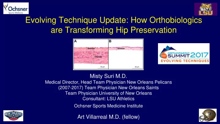
Evolving Technique Update: How Orthobiologics are Transforming Hip Preservation Misty Suri M.D. Medical Director, Head Team Physician New Orleans Pelicans (2007-2017) Team Physician New Orleans Saints Team Physician University of New Orleans
inner
Diseases of the Colon & Rectum 60 (6), E98-E98
2014;67(5):662-675.
– bone marrrow derived MSC adhesion – chondrogenic differentiation – chondrocyte adhesion & proliferation