1 4/17/2017 Cross section History Cornea Transplants Eye Recovery - - PowerPoint PPT Presentation

1
SMART_READER_LITE
LIVE PREVIEW

1 4/17/2017 Cross section History Cornea Transplants Eye Recovery - - PowerPoint PPT Presentation

4/17/2017 Minnesota Lions Eye Bank Mission Minnesota Lions Eye Bank Descemet Membrane Laboratory Department To help restore sight through eye donation Endothelial Keratoplasty (DMEK) Preparation Jeff Justin Clinical Laboratory Scientist


slide-1
SLIDE 1

4/17/2017 1

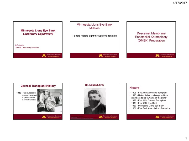
Minnesota Lions Eye Bank Laboratory Department

Jeff Justin Clinical Laboratory Scientist

Minnesota Lions Eye Bank Mission

To help restore sight through eye donation

Descemet Membrane Endothelial Keratoplasty (DMEK) Preparation

Corneal Transplant History

1905 First successful cornea transplant is performed in Czech Republic.

  • Dr. Eduard Zirm

Clinical & Experimental Ophthalmology Volume 33, Issue 6, pages 642-657, 5 DEC 2005 DOI: 10.1111/j.1442-9071.2005.01134.x http://onlinelibrary.wiley.com/doi/10.1111/j.1442-9071.2005.01134.x/full#f5

  • 1905 - First human cornea transplant
  • 1925 - Helen Keller challenge to Lions

members to be “Knights of the Blind”

  • 1937 - First U.S. Cornea Transplant
  • 1944 - First U.S. Eye Bank
  • 1960 - Minnesota Lions Eye Bank
  • 1961 - Eye Bank Association of America

History

slide-2
SLIDE 2

4/17/2017 2

  • 1974 < 5,000 cornea transplants
  • 2016 > 80,000 cornea transplants
  • Cornea transplants 95 % successful

History – Cornea Transplants

Cross section

  • Procurement - Enucleation

Eye Recovery Eye Recovery

  • Procurement - In Situ

Evaluation

  • Preserved cornea in viewing

chamber

Sclera

  • Whole sclera anchors

prosthetic eyes after enucleation.

  • Quarter sclera is used

to cover glaucoma shunts.

slide-3
SLIDE 3

4/17/2017 3

Specular Microscopy

Endothelial cell count

  • >2000/mm² for

endothelial transplant

  • Minimum cell density

needed for adequate function is ≈ 400−700 cells/mm².

Slit Lamp Biomicroscope

  • Scars
  • Edema
  • Defects
  • Guttae
  • Infiltrates
  • Foreign bodies

Ocular Coherence Tomography

  • Micron-resolution

images

  • Shows structural

features e.g., scars and defects

  • Allows precise

measurement of cornea dimensions

  • Used pre and post

processing

Pachymetry Suitability

  • Corneal condition determines which

surgery it’s suitable for:

– Penetrating Keratoplasty – Anterior Lamellar Keratoplasty – Deep Anterior Lamellar Keratoplasty – Endothelial Keratoplasties – Keratolimbal Allograft – Keratoprosthesis

Penetrating Keratoplasty (PK)

slide-4
SLIDE 4

4/17/2017 4

Penetrating Keratoplasty (PK) Superficial Corneal Scar Anterior Lamellar Graft (ALK) Keratolimbal Allograft (KLAL) for Stem Cell Deficiency

Cross section

Corneal endothelium

  • Purpose: control hydration (maintain

stromal deturgescence) and deliver nutrients and other molecules from the aqueous humor

  • 6000 cells/mm² at birth
  • 3400/mm² at age 15
  • 2300/mm² at age 85
slide-5
SLIDE 5

4/17/2017 5

  • DSAEK

Descemet Stripping Automated Endothelial Keratoplasty

  • DMEK

Descemet Membrane Endothelial Keratoplasty

Processed Tissue

Endothelial Keratoplasty Descemet’s Stripping Automated Endothelial Keratoplasty (DSAEK) Processing - DSAEK Microkeratome Dissection

Range of thickness for graft: 50-150 µm

Descemet’s Stripping Procedure Graft Insertion

slide-6
SLIDE 6

4/17/2017 6

Unfolding of graft DSAEK DMEK Loading the inserter Injecting graft Double scroll

slide-7
SLIDE 7

4/17/2017 7

Unfolding graft Unfolding graft Graft in place DMEK vs DSAEK

  • More anatomic with no optical interface
  • Better refractive outcomes/visual acuity
  • May have lower rejection rates
  • Faster recovery
  • Surgically more challenging as a

procedure

  • May have a higher dislocation rate

Descemets Membrane

  • Serves as the basement membrane of

the endothelial cells

  • Cells continually secrete collagen

throughout life

  • 3 µm thick at birth
  • 10-13 µm by age 70

Cornea suitability for DMEK

  • >50 yrs old− scroll not as tight & thicker
  • Endothelial cell count >2200 cells/mm²
  • No defects or scars affecting Descemets
  • Healthy endothelium
  • IOL scars- surgeon preference
  • Diabetes- ok
slide-8
SLIDE 8

4/17/2017 8

DMEK preparation

  • Secure on suction base
  • Score peripheral membrane
  • Free up edges
  • Peel membrane off stoma
  • Trephine and/or apply “S” stamp if

requested

Indications for DMEK/DSAEK

  • Similar for each
  • Post cataract surgery edema
  • Fuchs’ dystrophy
  • Repeat corneal transplant
  • Other causes of endothelial dysfunction
  • Unspecified

Tissue Utilization – US Eye Banks

Distribution 2016 2015 2014 2013 2012 Corneal Grafts Total 82,994 79,304 76,431 72,736 68,681 Penetrating keratoplasty 38,413 39,554 38,919 36,998 36,716 Anterior Lamellar Keratoplasty 2,386 2,201 1,953 2,009 1,855 Endothelial keratoplasty 32,221 30,710 28,961 27,298 24,277 Keratolimbal Allograft 97 107 88 110 97 Glaucoma Shunt Patch or

  • ther non keratoplasty use

917 527 755 687 676 Other keratoplasty 65 19 17 17 44 Unknown or Unspecified 1,514 1,142 1,026 1,068 1,554 Sclera 3,225 3,225 3,345 3,369 3,497 Research 17,023 16,924 17,670 17,384 19,320 Training 9,916 10,003 9,295 7,451 6,850

Domestic Endothelial Keratoplasty 2013 - 2016

Domestic Surgery Use 2016 2015 2014 2013 Total Endothelial Keratoplasty procedures 28,327 27,208 25,965 24,987 DSEK, DSAEK, DLEK Procedures 21,868 22,514 23,100 23,465 DMEK or DMAEK Procedures 6,459 4,694 2,865 1,522

Jeff Justin Laboratory Scientist JJJustin@umn.edu 612-625-3118

www.aGiftofSight.org

Information