
SLIDE 1
Embryology the anatomic basis of fetal medicine Prenatal consult - - PowerPoint PPT Presentation
Embryology the anatomic basis of fetal medicine Prenatal consult You meet with expectant parents and tell them that a congenital malformation has been identified. You proceed to explain the birth defect. Predictable questions Why
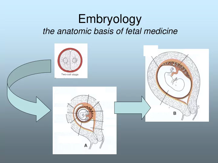
Monozygotic Twins
Completely separated after 2-cell stage – two chorionic cavities, two amniotic cavities. Separate uterine implantations
Monozygotic Twins
Separation of inner cell mass at later stages of development- resulting in common placenta – One chorionic cavity mono-chorionic A) separate amniotic cavities B) Single amniotic cavity A B
Dichorionic, diamniotic membranes Monochorionic diamniotic membranes Monochorionic Monoamniotic membranes
Which twins are at risk for Twin-twin Transfusion syndrome?
– Blastomeres / morula/ blastocyst – Trophoblast / embryoblast
– Endometrial embedding- development of placenta – Establishment of uteroplacental circulation by day 13 – Embryoblast – forms bilaminar germ disk and amniotic cavity lining develops
– Gastrulation – formation of 3 germ cell layers – Establishment of body axes
Events have to occur in correct spatial and correct time sequence
the Morula enters uterine cavity- and forms the blastocyst by day 9
1) Trophoblast (green) 2) Embryoblast ( blue/yellow)
Trimester 2 Trimester 1 Trimester 3
Day 12 – further embedding into endometrium
Trimester 2 Trimester 1 Trimester 3
Day 13: Established uteroplacental circulation Bilaminar disk stage
Future umbilical cord
Trimester 2 Trimester 1 Trimester 3
Development of tri-laminar disk Derivation of the three germ cell layers
Epiblast cells invaginate to form mesoderm
Trimester 2 Trimester 1 Trimester 3
Development of tri-laminar disk Derivation of the three germ cell layers
Establishment of body axes Looking onto ectoderm from above
Fate map for epiblast cells
pm: paraxial mesoderm= somites Im: intermed mesoderm= urogenital system, Lpm:lateral plate mesoderm= lateral body wall, eem:extraembryonic meso= chorion
Trimester 2 Trimester 1 Trimester 3
– Holoprosencephaly: – injury to anterior midline of germ disk- alcohol exposure / via SHH gene – Fusion of the eyes. – Single nasal chamber
– Caudal dysgenesis –
Example: 22-week fetus. The lower portion of the body is small compared with the midbody and chest. The lower extremities (arrows) appear abnormally extended and atrophied. Structures above the level of L3 and intracranial anatomy appear normal.
Source:radiology.rsnajnls.org/cgi/content/full/230/1/229
Day 28 of gestation Affects mesodermal derivatives ?lack of vascular supply? May be related to mat’l diabetes
– Situs inversus – – Generally autosomal recessive disorder – 5-10% have CHD most often transposition of the great vessels – If situs with levocardia (1in 2Mill) then 95% risk CHD – 25% will have primary ciliary dyskinesia (PCD) – 50% of PCD have Situs inversus= Kartagener syndrome siuts, sinusitis, bronchiectasis male infertility
– Sacrococcygeal tumors – arise from remnants of primitive streak.
Dermoid cyst or thyroglossal duct cyst?
Trimester 2 Trimester 3 Wk 4-8
Consequences of failure:……
Herniation of bowel through defect in abdominal wall – always to the right of umbilicus- Exposed intestine
Herniation through umbilical ring intestine covered by membrane
Question: Is it ever normal to see intestine
25 days 5 week embryo
hea
Which one(s) might you be able to dx prenatally?
Pulmonary agenesis- If bronchioles don’t grow- Lung parenchyma doesn’t grow
Pulmonary agenesis- If bronchioles don’t grow- Lung parenchyma doesn’t grow Cystic adenomatoid malformation Proliferation of bronchioles, not alveoli- abnormal sac of lung tissue
Pulmonary agenesis- If bronchioles don’t grow- Lung parenchyma doesn’t grow Cystic adenomatoid malformation Proliferation of bronchioles, not alveoli Pulmonary sequestration Separate piece of lung – not connected to Tracheobronchial tree Aortic blood supply
Pulmonary agenesis- If bronchioles don’t grow- Lung parenchyma doesn’t grow Cystic adenomatoid malformation Proliferation of bronchioles, not alveoli Congenital lobar emphysema Absent musculature on bronchus Results in hyperinflation Pulmonary sequestration Separate piece of lung – not connected to Tracheobronchial tree Aortic blood supply
Bronchogenic Cyst Diverticulum of tracheobronchial tree w/o associated pulmonary parenchyma
Bronchogenic Cyst Diverticulum of tracheobronchial tree w/o associated pulmonary parenchyma
Some are consequences of failures of normal developmental processes Some are accidents of nature when development has been fine
Some are consequences of failures of normal developmental processes Some are accidents of nature when development has been fine Does this make a difference in what you expect the incidence of associated anomalies to be????
Duodenal atresia/ stenosis: trisomy 21. cardiac defects, multiple atresias Jejunoileal atresia: No association with Genetic disorders or Other organ involvement
Di-chorionic, Di-amniotic membranes Mono-chorionic di-amniotic membranes Mono-chorionic Mono-amniotic membranes
Dizygotic twins 2 oocytes
Simultaneously fertilized – usually separate membranes, although they can fuse
– Conjoined twins – partial splitting of primitive node
http://library.med.utah.edu/WebPath/PEDHTML/PED022.html
– parenchymal development, retroperitoneal vs pelvic location, midline fusion
– Duplication anomalies, Insertion in bladder
– Size/innervation/musculature – Development of bladder neck, continence