
SLIDE 1
Property of VOMPTI, LLC For Use of Participants Only. No Use or Reproduction Without Consent 1
www.vompti.com
Orthopaedic Manual Physical Therapy Series 2017-2018
Orthopaedic Manual Physical Therapy Series Charlottesville 2017-2018
ELBOW ANATOMY, BIOMECHANICS
AND PATHOLOGY
Kristin Kelley, DPT, OCS, FAAOMPT
Orthopaedic Manual Physical Therapy Series 2017-2018 www.vompti.com
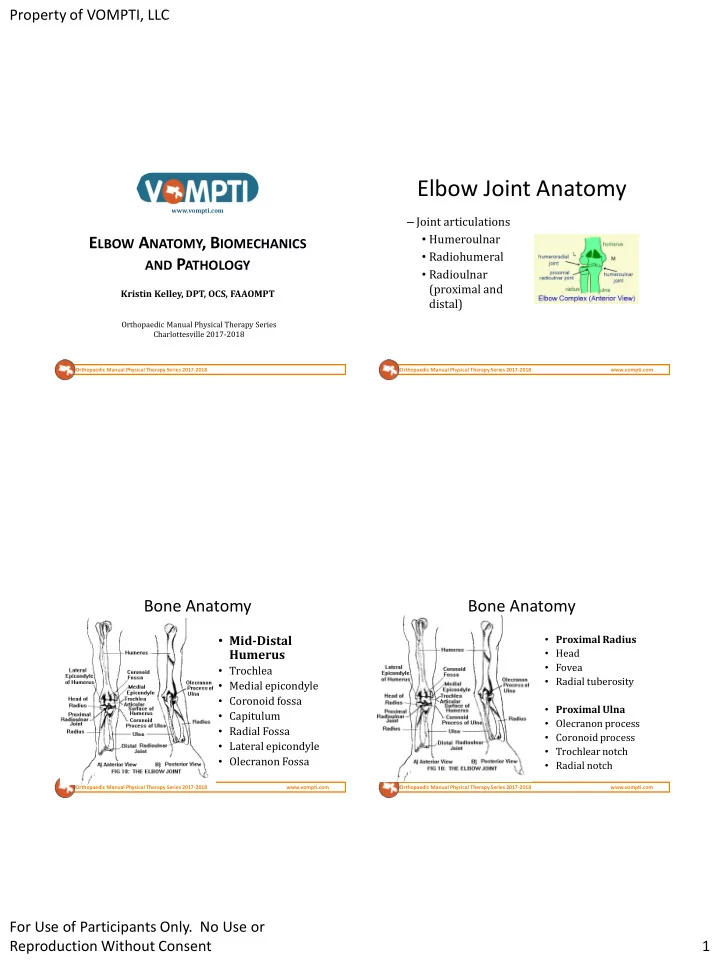
Elbow Joint Anatomy
– Joint articulations
- Humeroulnar
- Radiohumeral
- Radioulnar
(proximal and distal)
Orthopaedic Manual Physical Therapy Series 2017-2018 www.vompti.com
Bone Anatomy
- Mid-Distal
Humerus
- Trochlea
- Medial epicondyle
- Coronoid fossa
- Capitulum
- Radial Fossa
- Lateral epicondyle
- Olecranon Fossa
Orthopaedic Manual Physical Therapy Series 2017-2018 www.vompti.com
Bone Anatomy
- Proximal Radius
- Head
- Fovea
- Radial tuberosity
- Proximal Ulna
- Olecranon process
- Coronoid process
- Trochlear notch
- Radial notch