SLIDE 1
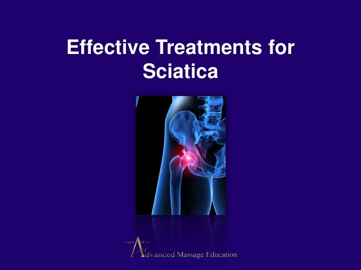
Effective Treatments for Sciatica
SLIDE 2 Exact data on the incidence and prevalence of sciatica are lacking. In general an estimated 5%-10% of patients with low back pain have sciatica, whereas the reported lifetime prevalence of low back pain ranges from 49% to 70%. The annual prevalence of disc related sciatica in the general population is estimated at 2.2% Most patients with acute sciatica have a favorable prognosis but about 20%-30% have persisting problems after one or two years. The diagnosis of sciatica and its management varies considerably within and between countries. Disc surgery may provide quicker relief of leg pain than conservative care but no clear differences have been found after one or two years.
SLIDE 3
Gluteal and Sciatic Nerves
SLIDE 4
SLIDE 5
SLIDE 6
SLIDE 7
Acland’s DVD Atlas of Human Anatomy The Lower Extremity Lippincott Williams and Wilkins
SLIDE 8 Most acute back problems are self- limiting:
- Around 90% will be better within 6 weeks
regardless of the treatment they receive.
- Bed rest is almost never of value for non-
specific pain.
- Between 2% and 8 % will develop chronic
symptoms.
- Most people can point to a single event.
SLIDE 9 Carol Hartigan, MD
Fear-avoidance behavior and Reduced activity levels are common. 'Let's not guard it and protect it anymore, It's the opposite
- f what they have been told.
If you have a bad back, it should be strong and flexible and fit."
Assistant Clinical Professor, Physical Medicine & Rehab, Harvard Medical School
SLIDE 10
The Spine Center at New England Baptist Hospital SpineCenter@NEBH.org
Rainville J, Hartigan C, et al. 2004. Exercise as a treatment for chronic low back pain. The Spine Jour 4: 106-115.
Carol Hartigan , MD
SLIDE 11 Vladimir Janda, MD
Is best known for his identification of the upper crossed; and lower crossed syndromes. Each of these clinical scenarios described conditions in which the tone of antagonistic muscle groups became imbalanced and led to the predictable sequence of pain and dysfunction.
Key figure in the 20th Century rehabilitation movement
SLIDE 12
Founder of the Muscular Therapy Institute in Cambridge, MA.
The low back is the most commonly injured part of the body. The most common low back injury is the Sacroiliac ligaments.
SLIDE 13
benbenjamin.com
benbenjamin.com.au
Ligaments of the Sacrum: The Primary Cause of Low Back Pain
benbenjamin.com/pdfs/08so.pdf
SLIDE 14
SLIDE 15
SLIDE 16
SLIDE 17
SLIDE 18
Prolotherapy
SLIDE 19
SLIDE 20
Ross A. Hauser M.D. drhauser@caringmedical.com
www.caringmedical.com Oak Park, IL 60301
SLIDE 21
SLIDE 22
SLIDE 23
SLIDE 24 Contributing Factors
- Prolonged sitting - The single most stressful
event in the low back which puts all of the weight
- f the upper body on the sacroiliac area.
- This posture can lead to tight hip flexor muscles
that encourage weakness in the low back.
If no violent falls or traumas have occurred, many contributing factors can play a large part in damaging the sacroiliac region and supporting ligaments.
SLIDE 25 Contributing Factors
- Bending of the knees in this position will
cause tight hamstrings muscles which pull the ischium downwards that further strains the low back.
- Poor posture - Will magnify strain on the
vertebral discs and sacral ligaments.
SLIDE 26 Contributing Factors
Poor posture habits are sometimes ingrained from an early age. Many causes include: being tall in height as a
- child. Not being encouraged to stand up straight.
Wearing shoes with unusually high heel heights.
SLIDE 27
Contributing Factors Inadequate support - Weak abdominal muscles do not allow the spine to stay balanced evenly on the pelvis causing more pronounced spinal curvature. Relaxing in soft furniture will eventually influence posture, which weakens supporting structures of the spine.
SLIDE 28
SLIDE 29
Contributing Factors Poor flexibility - The body responds to poor flexibility by altered or limited mobility. This change in mobility results in unnecessary stress on the joints, poor circulation, and weakness in other muscle groups.
SLIDE 30 Dissecting Low Back Pain
Before the musculature of the low back can be examined we must look at the movements we should be capable of.
SLIDE 31
SLIDE 32 Assessment is the key
A series of question and answers: When did you first notice the problem? Can a certain movement reproduce any pain? If you used any previous treatment, was it helpful? Any muscle aches, tension, or problems sleeping? Be sure to address the list of contributing factors in the previous chapter. Encourage the person to make lifestyle changes if many of the factors are on their list.
In order the effectively treat sacroiliac joint dysfunction, or lower body pain assessment skills must be practiced and perfected.
SLIDE 33
SLIDE 34
Functional Assessment Protocol
Resistance or pressure from the therapist is only 1 to 2 pounds. Direction of resistance follows black arrows on illustration. Test is performed for a maximum of 5 seconds.
SLIDE 35 Trunk Extension
- Position of the Subject: Prone with head and
upper trunk extending off the table from about the nipple line. Arms at sides.
- Position of Therapist: Standing at side of table.
Lower extremities are stabilized just above the ankles.
- Test: Subject extends spine, raising body from the
table so that the umbilicus clears the table.
- Instructions to Subject: “Raise your head, arms,
and chest from the table as high as you can”.
SLIDE 36 Trunk Flexion
- Position of Subject: Supine with hands clasped behind head. Grade 3, arms
- utstretched in full extension above plane of body.
- Position of Therapist: Standing at side of table. Lower extremities are
stabilized just above the ankles.
- Test: Subject flexes trunk through range of motion. A curl up is emphasized,
and trunk is curled until scapulae clear table.
- Instructions to Subject: “Tuck your chin
and bring your head, shoulders, and arms off the table, as in a sit-up”
SLIDE 37 Trunk Rotation
- Position of Subject: Supine with hands clasped behind head.
- Position of Therapist: Standing at side of table. Lower extremities are
stabilized just above the ankles.
- Test: Subject flexes trunk and rotates to one side. This movement is then
repeated on the opposite side.
- Instructions to Subject: “Lift your head and shoulders from the table, taking
your right elbow toward your left knee.” Reverse.
SLIDE 38 Hip Extension
- Position of Subject: Prone.
- Position of Therapist: Standing next to limb. Hand to
give 1 to 2 pounds of resistance is placed on the posterior leg just above the ankle. The opposite hand may be used to stabilize pelvis at the Sacrum.
- Test: Subject extends hip. Resistance is given straight
downward to the floor.
- Instructions to Subject: “Lift your leg off the table
without bending your knee and don’t let me push it down.”
SLIDE 39 Hip Flexion
- Position of Subject: Sitting on table with thighs fully supported and lower legs
hanging over the edge.
- Position of Therapist: Standing next to limb.
The contoured hand to give 1 to 2 pounds of resistance over distal thigh just proximal to the knee.
- Test: Subject flexes hip, clearing the table.
- Instructions to Subject: “Lift your leg off
the table and don’t let me push it down.”
SLIDE 40 Hip External Rotation
- Position of Subject: Sitting on table, supported by hands.
- Position of Therapist: Kneels beside limb to be tested. The hand that
gives resistance grasps the ankle. 1 to 2 pounds of resistance is applied as a laterally directed force at the ankle. The other hand which offers counter pressure is contoured over the lateral thigh just above the knee. Resistance of 1 to 2 pounds is given as a medially directed force at the knee.
- Test: Subject externally rotates the
hip.
- Instructions to Subject: “Don’t
let me turn your leg out.”
SLIDE 41 Hip Adduction
- Position of Subject: Side-lying with test limb resting on the table. Uppermost
limb is supported by therapist by cradling the leg with the forearm.
- Position of Therapist: Standing behind subject. The hand giving resistance to
the lowermost limb is placed on the medial distal part of the femur.
- Test: Subject adducts hip until the lower limb contacts the upper one. Using 1 to
2 pounds of resistance.
- Instructions to Subject: “Lift your bottom leg up to your top one. Hold it. Don’t
let me push it down.”
SLIDE 42 Hip Abduction
- Position of Subject: Side-lying with test leg uppermost.
Lowermost leg is flexed for stability.
- Position of Therapist: Standing behind subject. The hand
giving resistance of 1 to 2 pounds is contoured across the lateral surface of the knee. The other hand is used to palpate Gluteus medius and minimus.
- Test: Subject abducts hip without
flexing the hip.
- Instructions to Subject: “Lift your leg up
in the air. Hold it. Don’t let me push it down.”
SLIDE 43
Assessment of Pelvic Girdle
SLIDE 44
Posterior legs – Gastrocnemius
SLIDE 45
Posterior legs – Hamstrings
SLIDE 46
SLIDE 47
Low back – Lateral rotator tendons
SLIDE 48
Low back – Lateral rotator tendons
SLIDE 49
Low back - Sacral ligaments
SLIDE 50
Low back - Sacral ligaments
SLIDE 51
Low back - Iliolumbar ligaments
SLIDE 52
SLIDE 53
Low back - Sacroiliac fascia
SLIDE 54
Low back - Sacroiliac fascia
SLIDE 55
Lateral Raffe – Quadratus Lumborum
SLIDE 56
SLIDE 57
SLIDE 58 Cadaver Dissection Series: Low Back Middle layer of Thorcolumbar Fascia, Lateral Raffe/Lateral Abdominal Wall. ECI Inc. 1303 Marsh Lane, Carrollton,
SLIDE 59 Posterior lumbar fascial compartment surrounded by the posterior and middle layer of the lumbar fascia. The posterior layers originate medially from the lumbar spinous processes and interspinous ligaments and wrap around laterally to join the lateral raphe, the dense union where the posterior and middle layers meet. The middle layer provides the fascial anterior border of the erector spinae muscles as it attaches to the tips of the lumbar transverse processes and is directly continuous with the intertransverse ligaments.
SLIDE 60
Treatment of a Case of Subacute Lumbar Compartment Syndrome Using the Graston Technique by Warren I. Hammer, DC, MS
SLIDE 61
Mid back – Erector Spinae
SLIDE 62
Anterior legs – Quadriceps
SLIDE 63
Anterior legs – Quadriceps
SLIDE 64
Anterior hip - Inguinal ligament
SLIDE 65
Active isolated assisted-stretching
These movements will not only elongate tight muscles and ligaments that you have just released, reeducate the injured tissue, but will also strengthen weaken areas without activating the stretch reflex.
SLIDE 66
Posterior Calf stretch (prone)
SLIDE 67
Hamstring stretch (supine)
SLIDE 68
Sacroiliac fascia stretch (passive movement supine)
SLIDE 69
Erector Spinae Stretch (passive movement standing)
SLIDE 70
Quadratus Lumborum stretch (passive movement supine)
SLIDE 71
Quadriceps stretch – (prone)
SLIDE 72
Inguinal ligament stretch (passive side lying)
SLIDE 73
Quadriceps/Hip flexor stretch (additional)
SLIDE 74
Kinesiology Tape Treatment
SLIDE 75 Self treatment
Other than the treatment you have given, the person’s best chance of success with their pain is realizing that the management of their back is their responsibility. Self treatment will be more effective in the long term management of their pain than any other form of treatment.
Encourage the person to be as active as possible even if they are experiencing pain. Awareness of improper posture and self stretches will reeducate the lower body, retard scar tissue formation and will continue to improve range of motion.
SLIDE 76
SLIDE 77
AdvancedMassageEducation.com